- 2
- ,
- 3
- 2
- 7
To Quiz Yourself: Select OFF by clicking the button to hide the diagnosis & additional resources under the case.
Quick Browser: Select ON by clicking the button to hide the additional resources for faster case review.
CASE NUMBER
310


Diagnosis
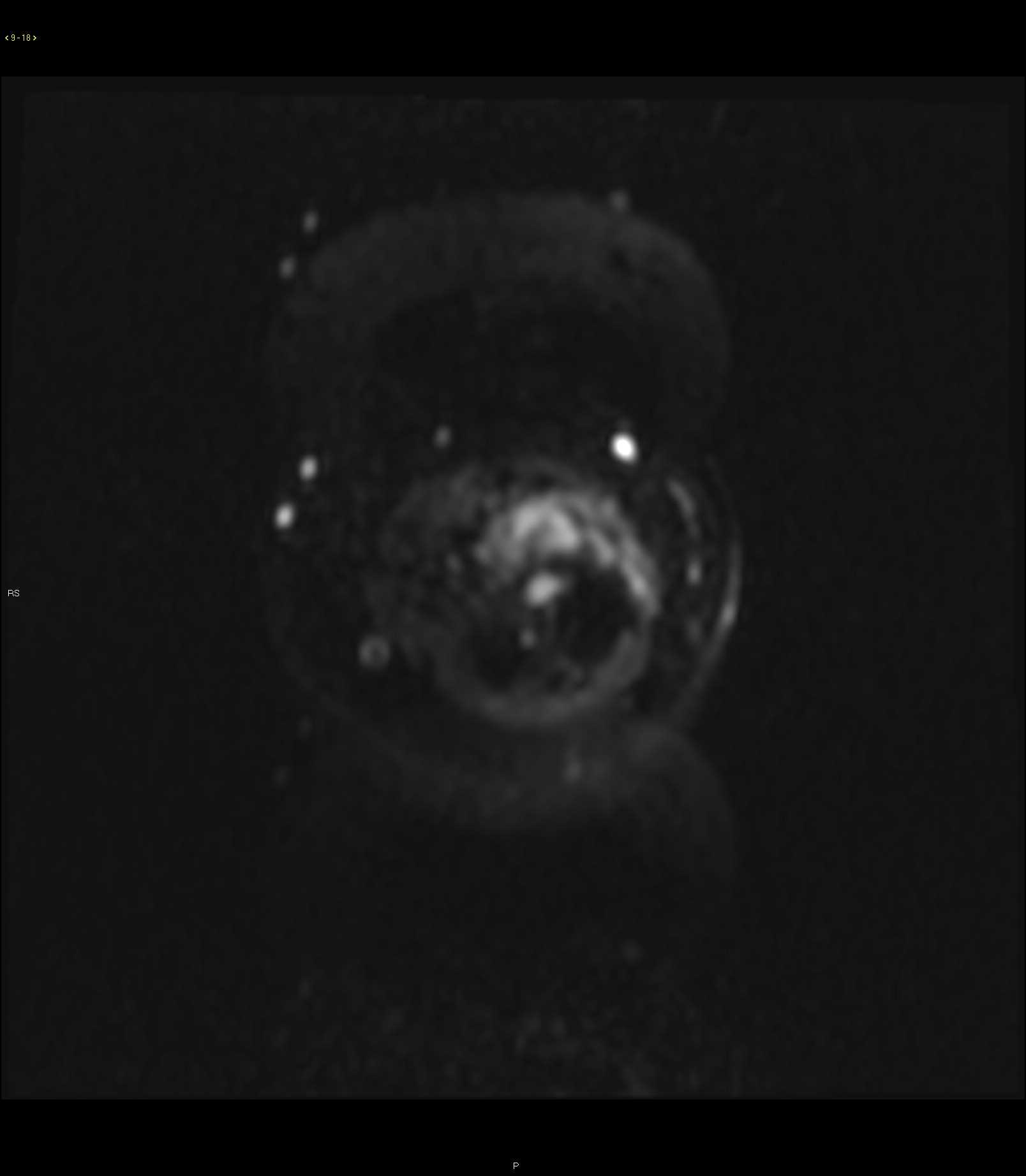
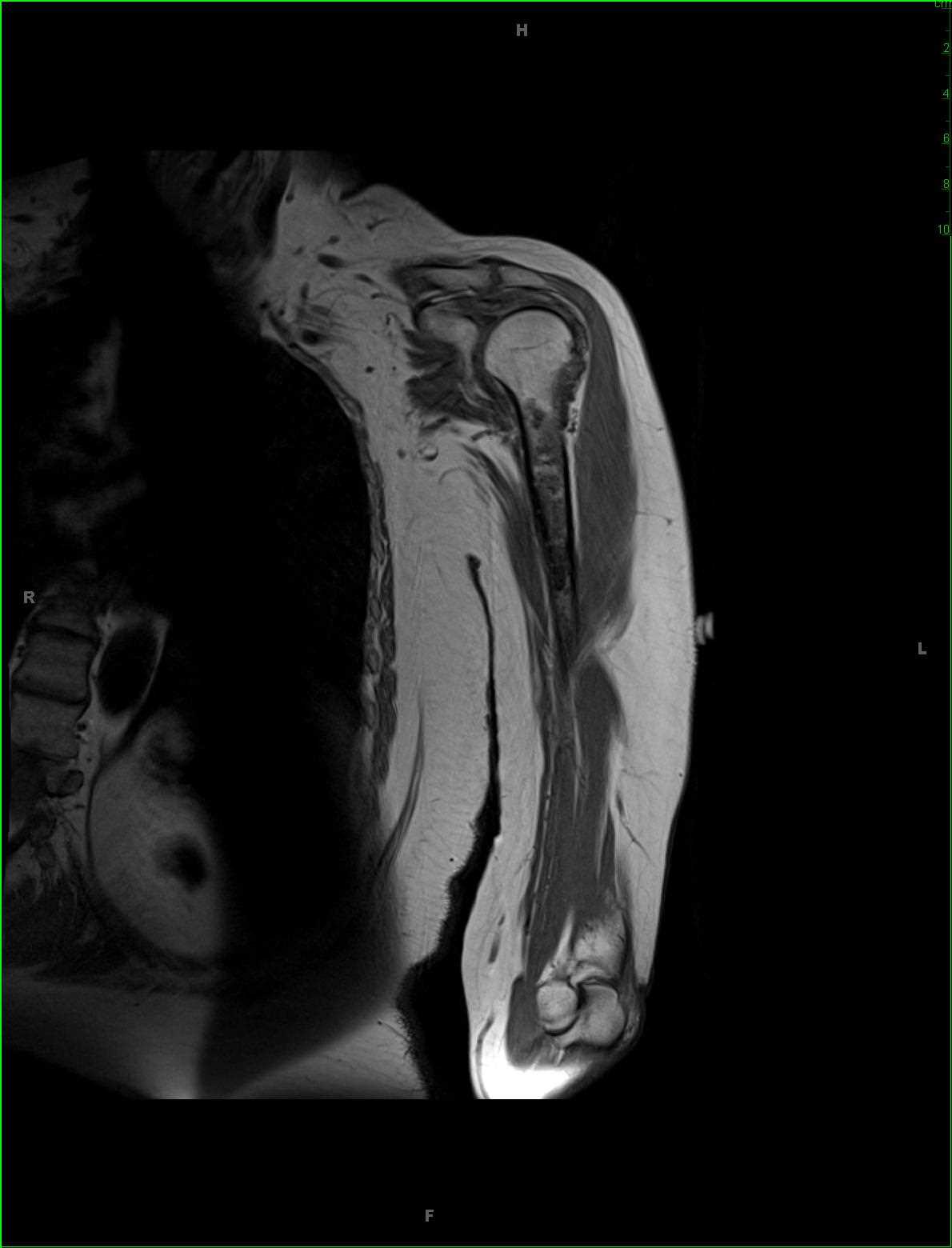
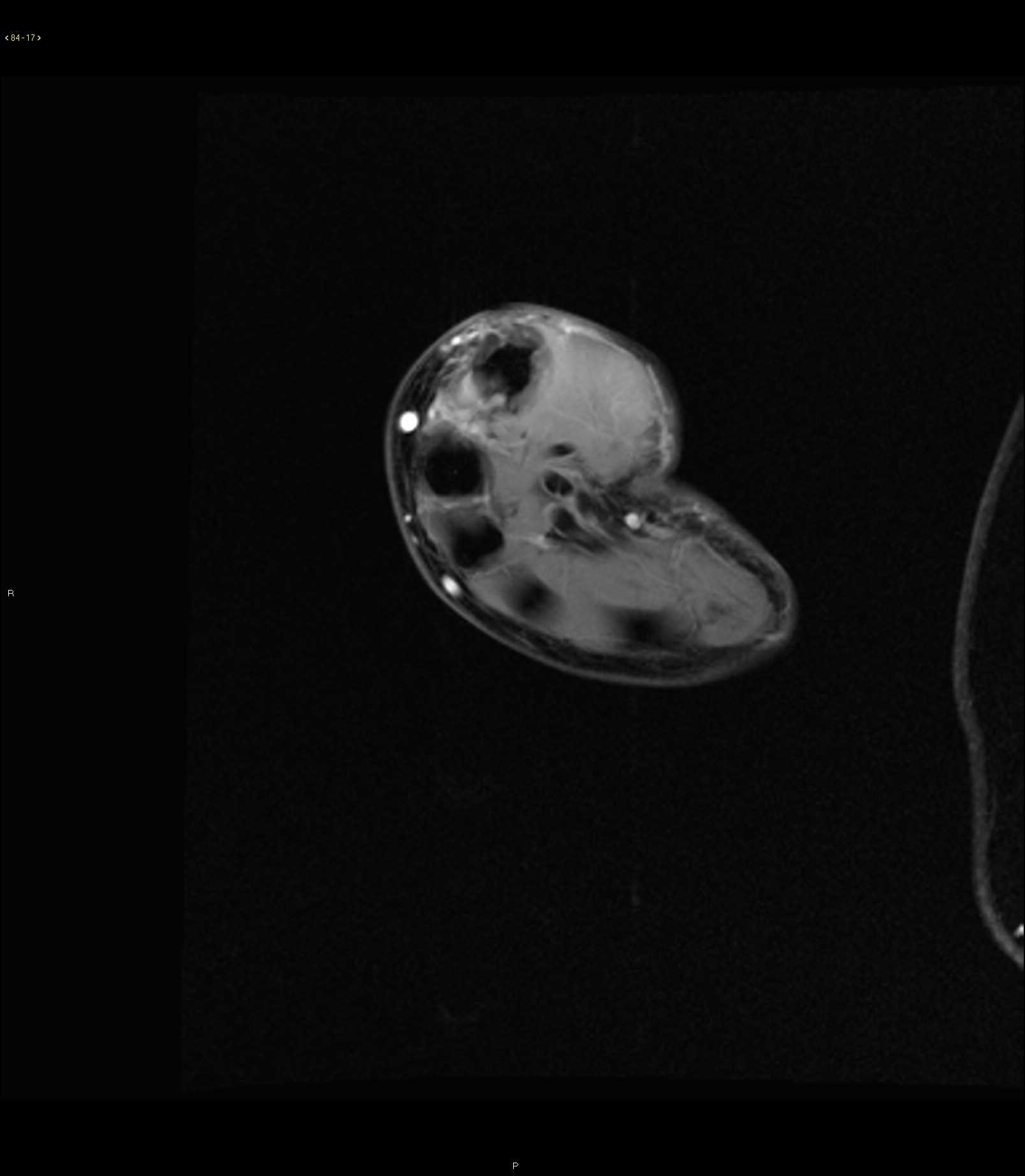
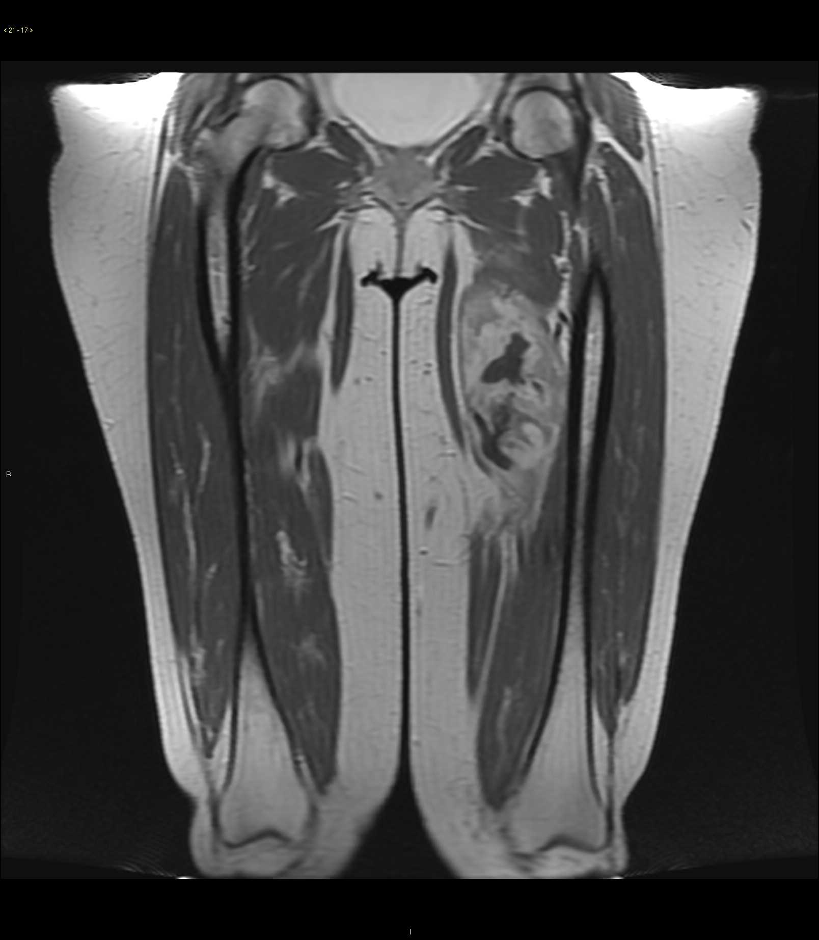
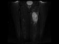
Myxofibrosarcoma grade 3
Note
45-year-old male with a slowly enlarging left thigh mass. There is a well-circumscribed solid appearing T1 mildly heterogeneous but predominantly isointense, T2/STIR heterogeneously hyperintense, centrally necrotic peripherally enhancing mass in the mid medial left thigh. A small plane of fat is maintained between the lateralmost lesion boundary and the cortical margin of the left femoral diaphysis at that site. There is mild perilesional edema. A myxoid tumor of the soft tissues was favored. This lesion was found to be a grade 3 myxofibrosarcoma. Imaging features of myxoid soft tissue tumors include high signal intensity characteristics on T2 weighted images because of high water content. Lesions range from benign to malignant entities. Some of the benign etiologies include intramuscular myxoma, synovial cyst, bursa, ganglion and benign peripheral nerve sheath tumor. Malignant etiologies include myxoid leiomyosarcoma, myxoid liposarcoma, myxoid chondrosarcoma and myxoid fibrosarcoma. Treatment for the malignant subtypes is usually with preoperative chemoradiation therapy followed by gross total resection.
Related videos to the case

THIS IS CASE
310
OF
370