- 2
- ,
- 3
- 2
- 7
To Quiz Yourself: Select OFF by clicking the button to hide the diagnosis & additional resources under the case.
Quick Browser: Select ON by clicking the button to hide the additional resources for faster case review.
CASE NUMBER
208


Diagnosis
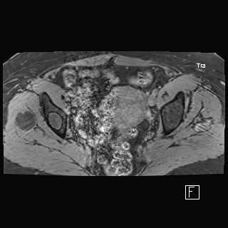
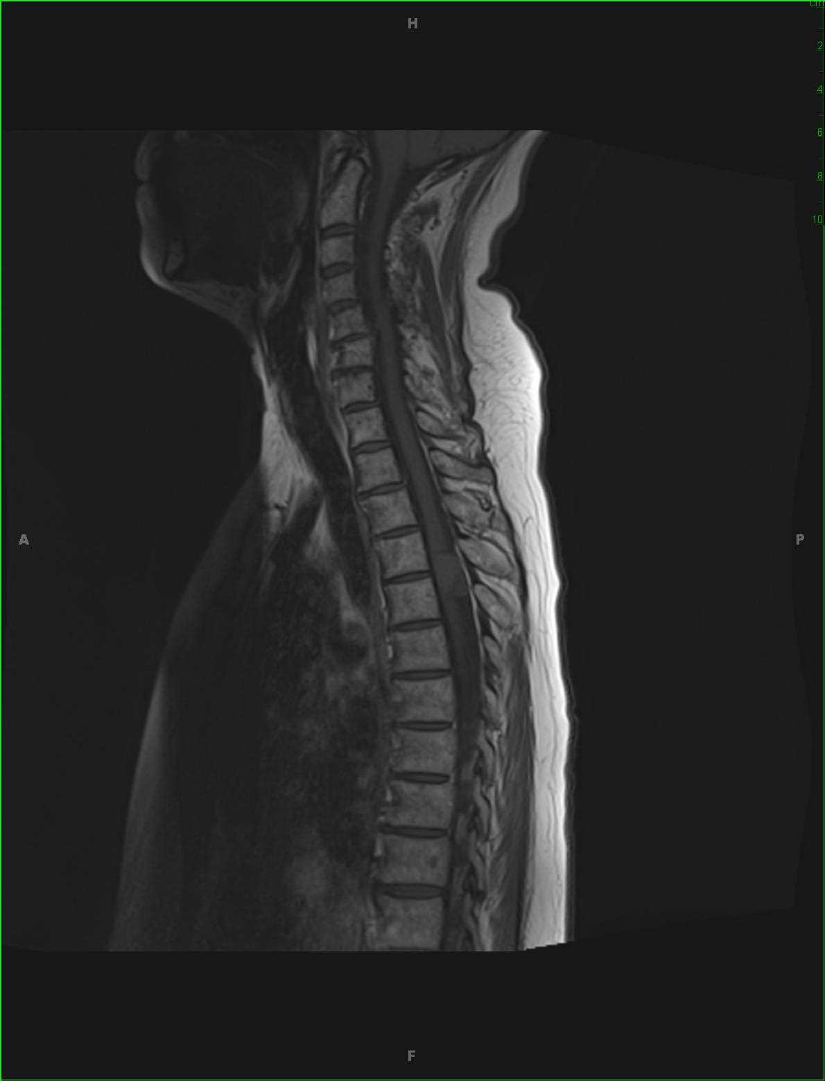
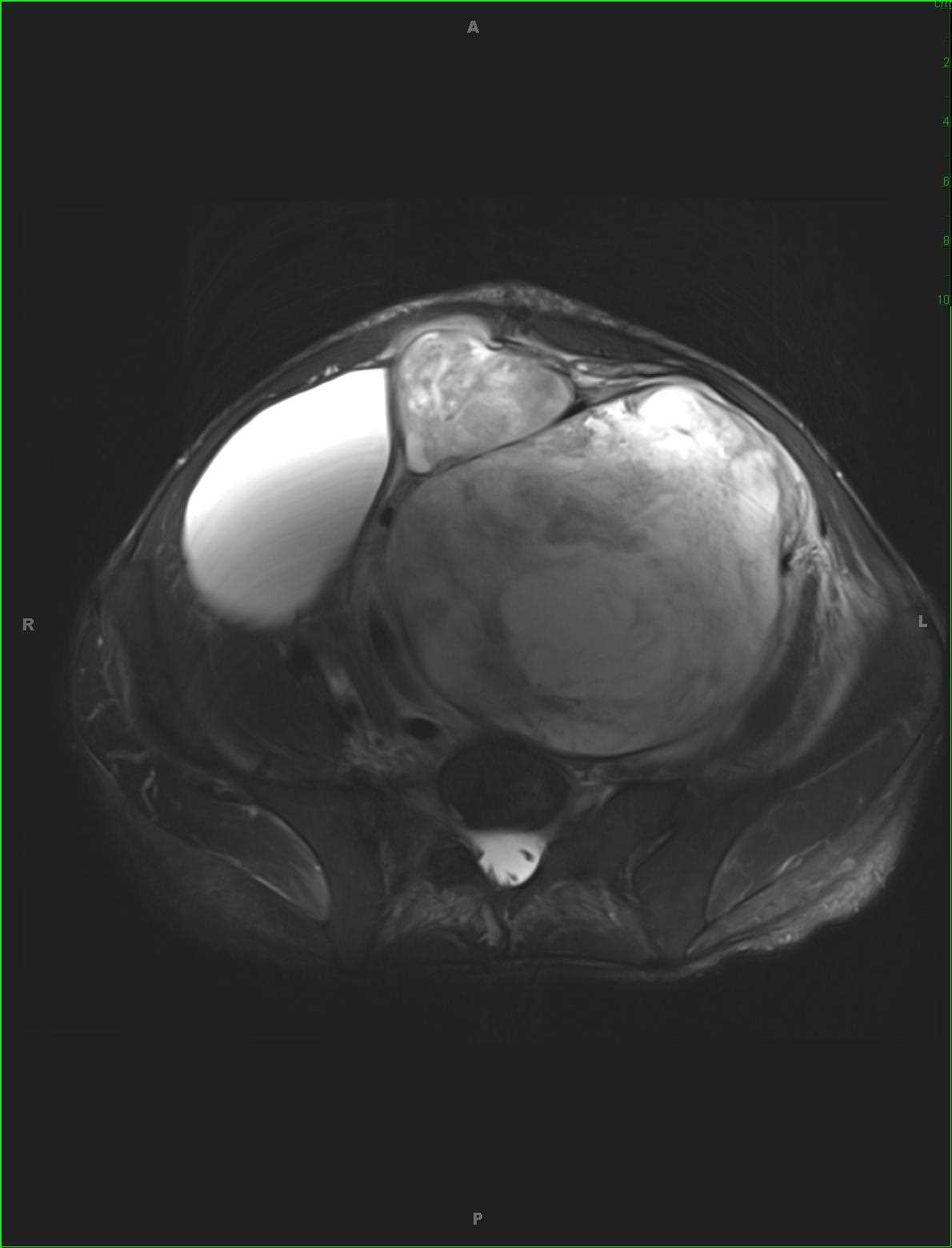
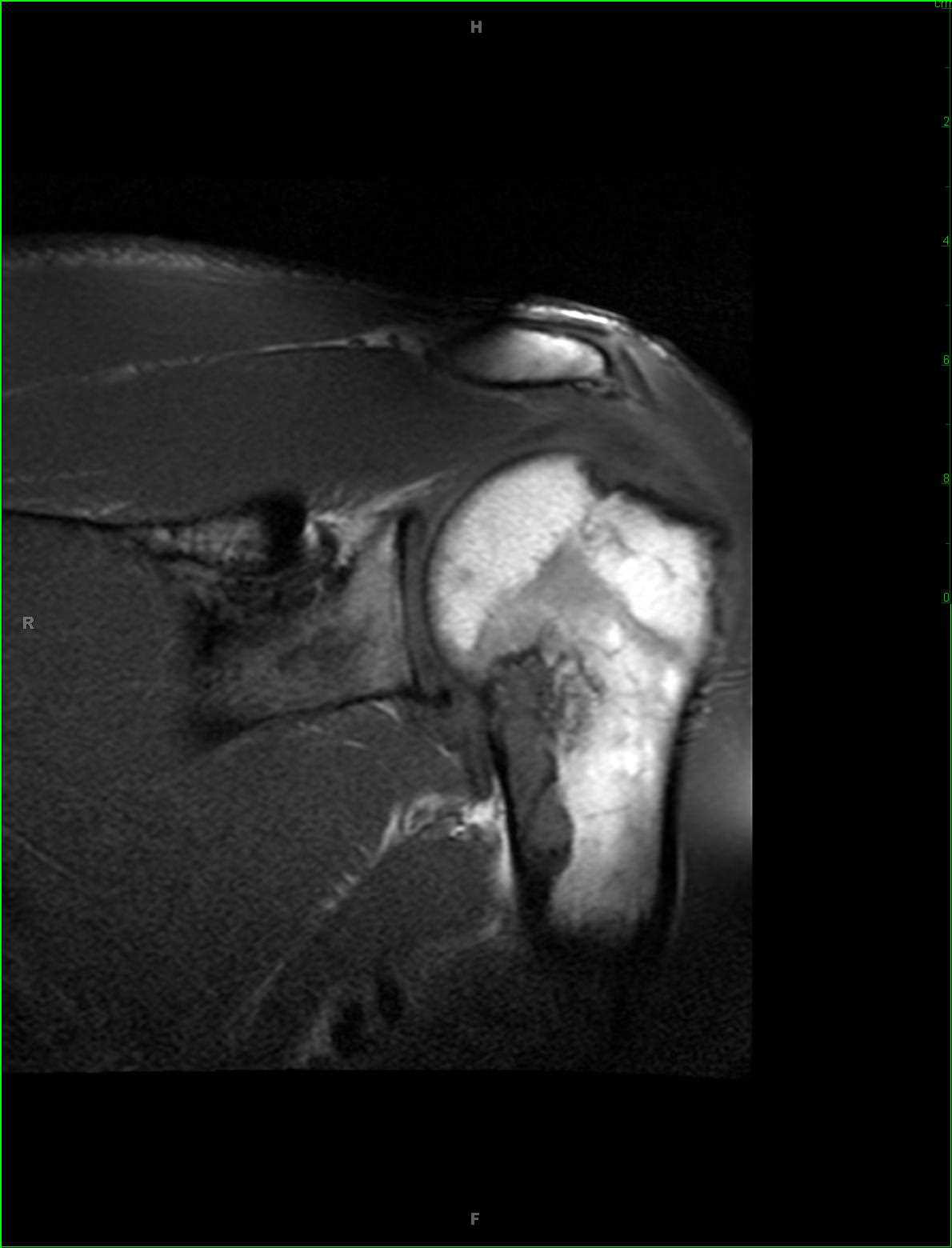
Polyostotic Fibrous Dysplasia
Note
Centered within the proximal left humeral diaphysis, there is an eccentrically located, cortically based T1-hypointense lesion with lobular margins. On the axial GRE weighted images, additional lesions are identified within the glenoid neck. The lesions are T2/STIR hyperintense and heterogeneously enhance. Imaging findings are classic for polyostotic fibrous dysplasia. Fibrous dysplasia is a non-neoplastic tumor like congenital process resulting in a localized absence of osteoblast differentiation maturation with replacement of normal bone with large fibrous stroma and islets of immature woven bone. The disease occurs predominantly in children and young adults. In the polyostotic form, patients are usually diagnosed by 10 years of age. The osseous structures affected by fibrous dysplasia are weaker than normal bone is susceptible to pathologic fracture. Sarcomatous degeneration to osteosarcoma, fibrosarcoma, chondrosarcoma or malignant fibrous histiocytoma may occasionally be seen.
Related videos to the case


THIS IS CASE
208
OF
370