- 2
- ,
- 3
- 2
- 7
To Quiz Yourself: Select OFF by clicking the button to hide the diagnosis & additional resources under the case.
Quick Browser: Select ON by clicking the button to hide the additional resources for faster case review.
CASE NUMBER
201


Diagnosis
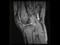
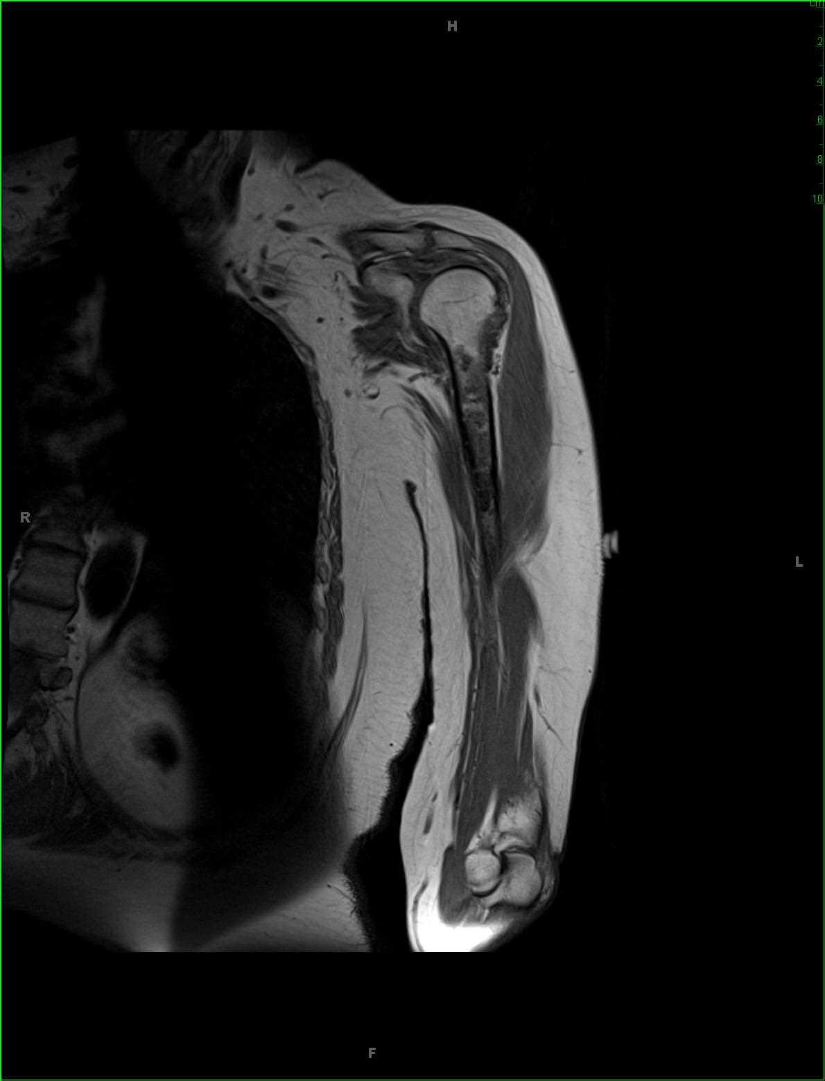
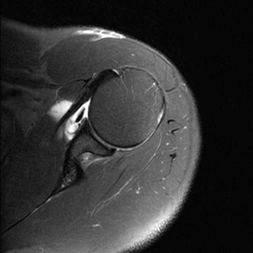
Pigmented Villonodular Synovitis, Knee
Note
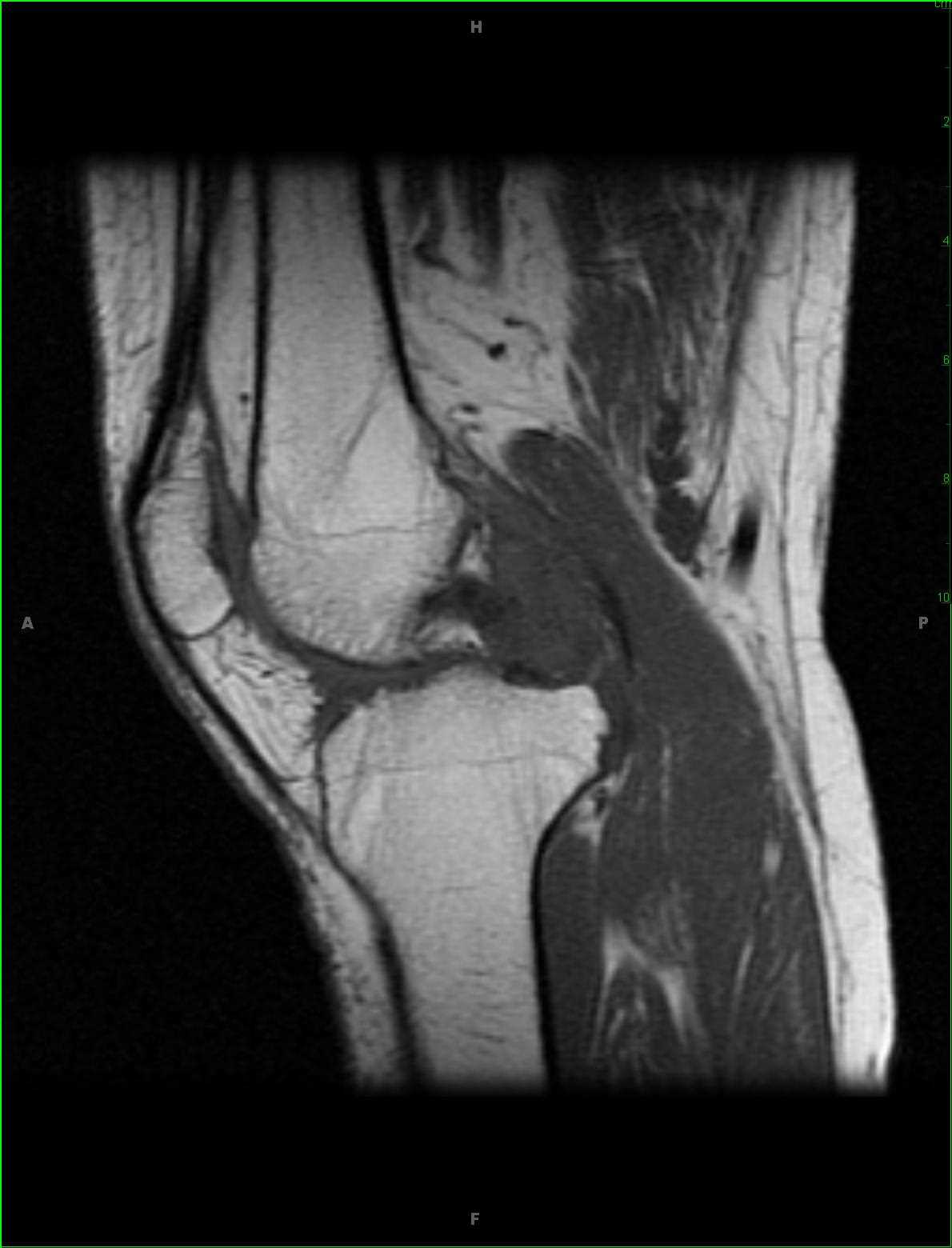
35-year-old male with chronic left knee pain. The images demonstrate a proton density isointense, STIR mildly hypointense lesion with scattered regions of signal loss. The lesion heterogeneously enhances and is intimately associated with the posterior cruciate ligament. These imaging findings are characteristic of pigmented villonodular synovitis or PVNS for short. PVNS is a benign proliferative condition affecting the synovial membranes of joints, bursa, or tendons. It results from neoplastic synovial proliferation with villous and nodular projections with scattered hemosiderin deposition. It is most common monoarticular. Occasionally, PVNS can look like aggressive neoplasms such as rhabdomyosarcoma, synovial sarcoma, or epithelial sarcoma.
Related videos to the case