- 2
- ,
- 3
- 2
- 7
To Quiz Yourself: Select OFF by clicking the button to hide the diagnosis & additional resources under the case.
Quick Browser: Select ON by clicking the button to hide the additional resources for faster case review.
CASE NUMBER
200


Diagnosis
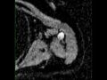
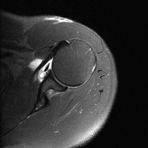
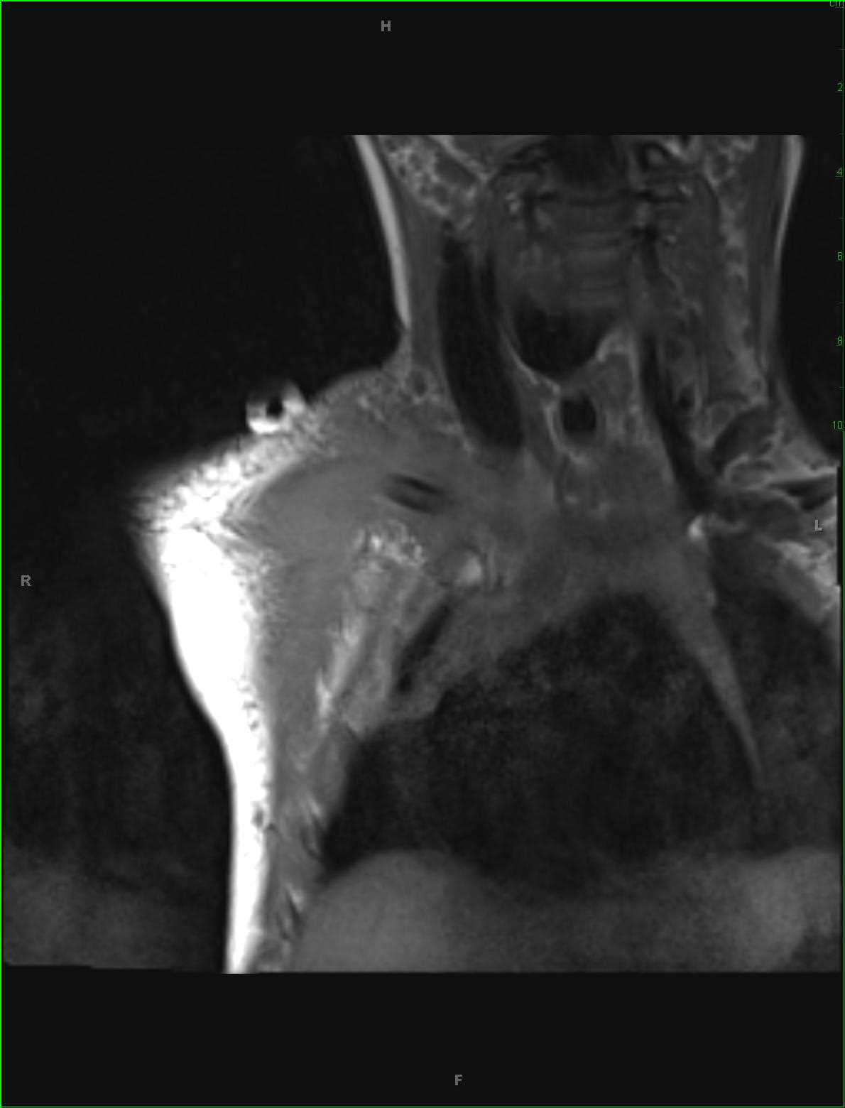
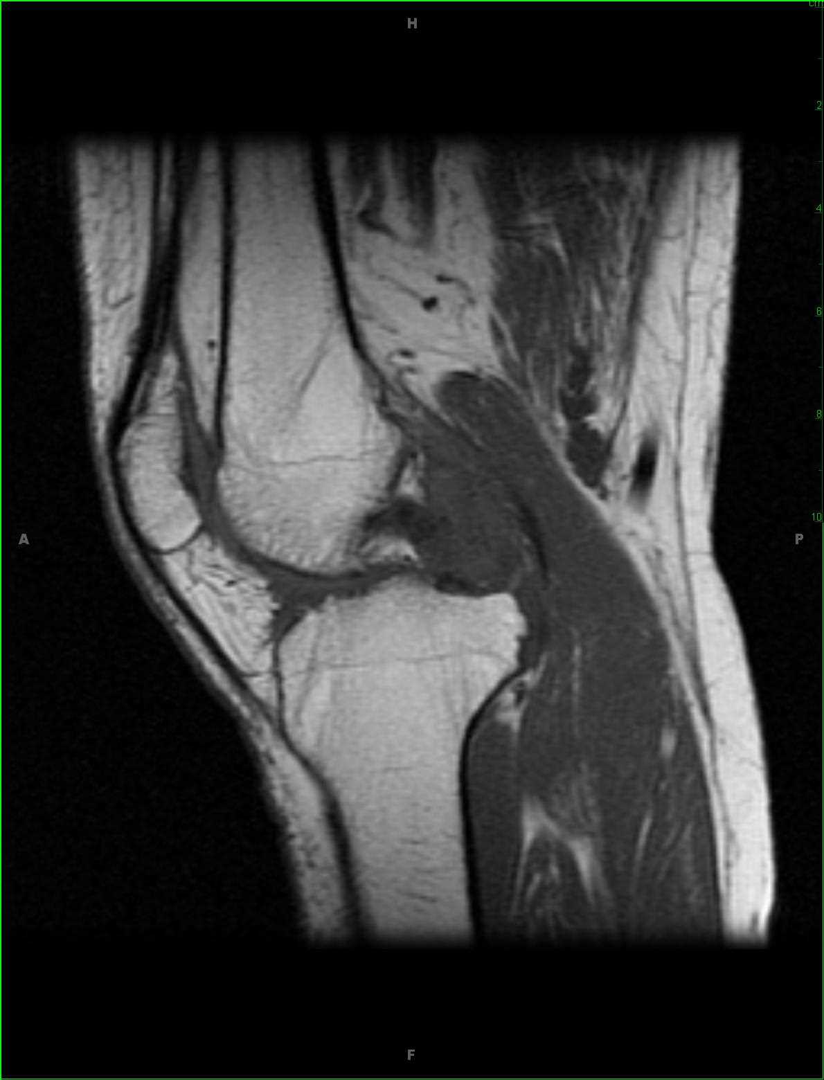
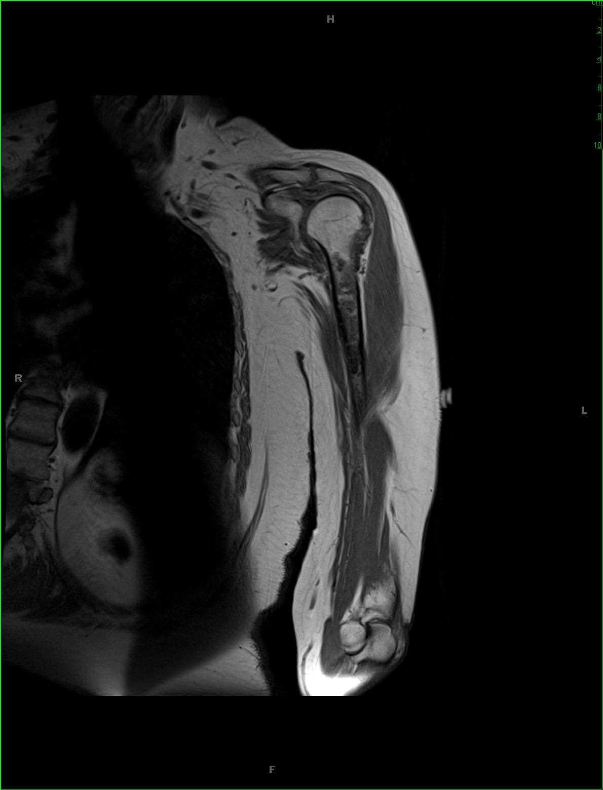
Conventional Chondrosarcoma, Humerus
Note
58-year-old female with history of chronic left arm pain. There is a T1 hypointense, STIR heterogeneously hyperintense lesion centered within the medullary cavity of the left humeral diaphysis. There are nodular regions of diffusion restriction with heterogeneous enhancement. There is subtle endosteal scalloping. The imaging characteristics are those of a chondroid lesion. Given the overall size and appearance, conventional chondrosarcoma was favored and was found on biopsy. The differential also included enchondroma and much less likely bone infarct. Conventional chondrosarcoma is the most common subtype. They typically occur in the fourth and fifth decades of life with a slight male predominance. At time of diagnosis, the lesion is usually over 4 cm in diameter. Typical clinical presentations include pain, palpable mass, and pathologic fracture.
Related videos to the case