- 2
- ,
- 3
- 0
- 9
To Quiz Yourself: Select OFF by clicking the button to hide the diagnosis & additional resources under the case.
Quick Browser: Select ON by clicking the button to hide the additional resources for faster case review.
CASE NUMBER
59
Diagnosis
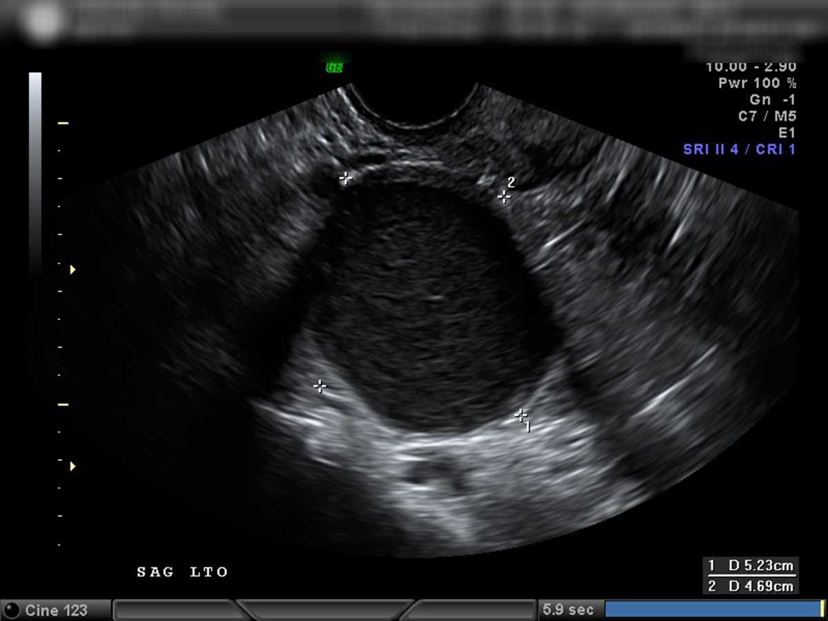
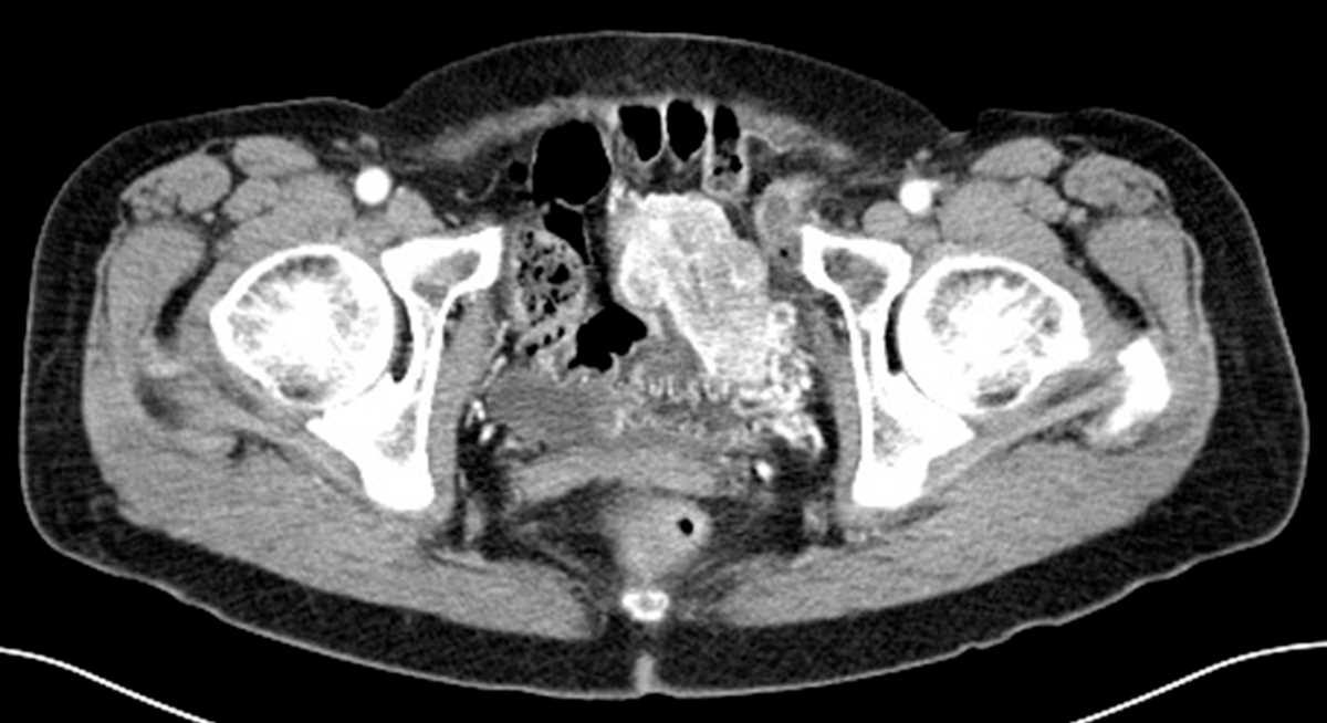
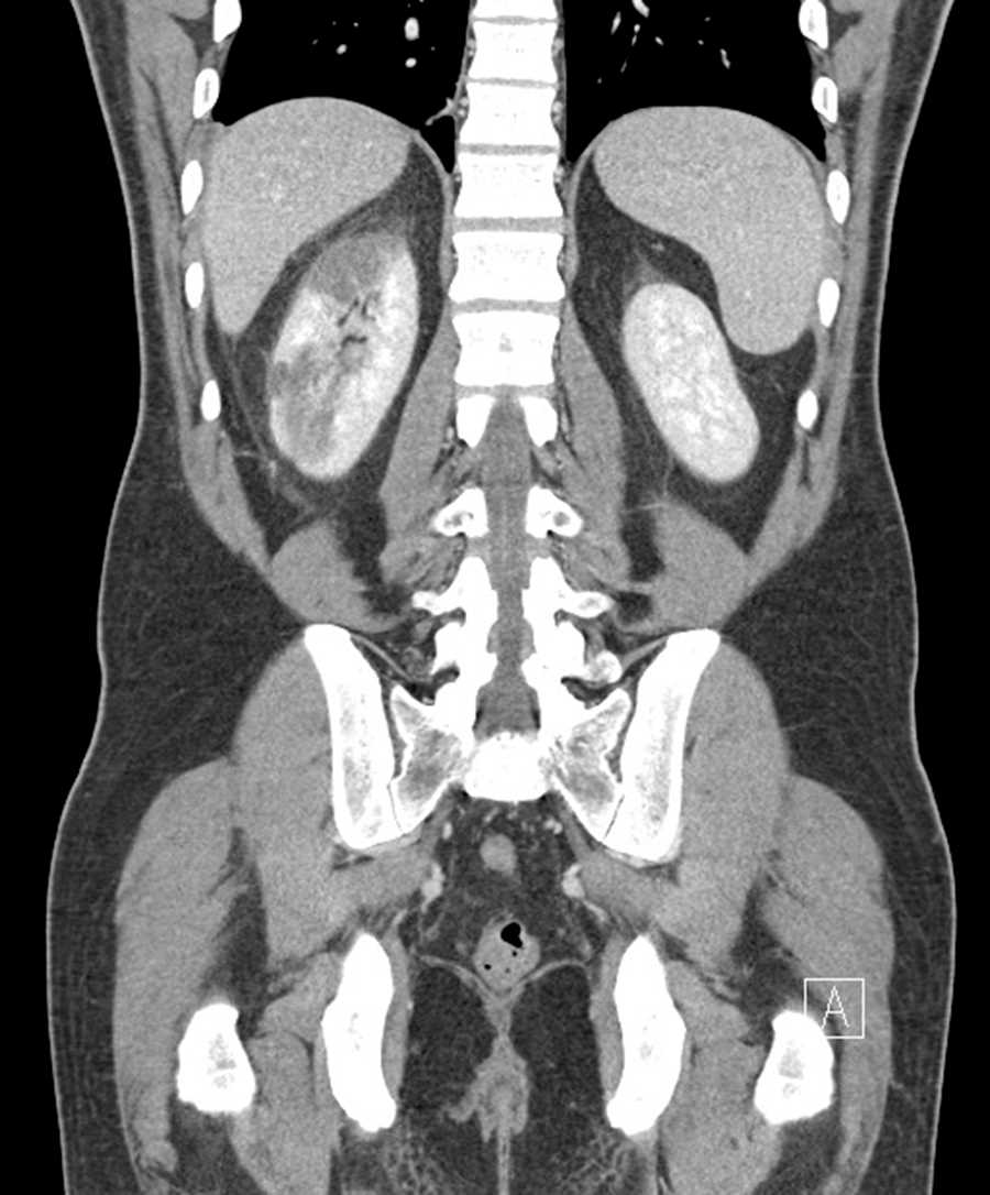
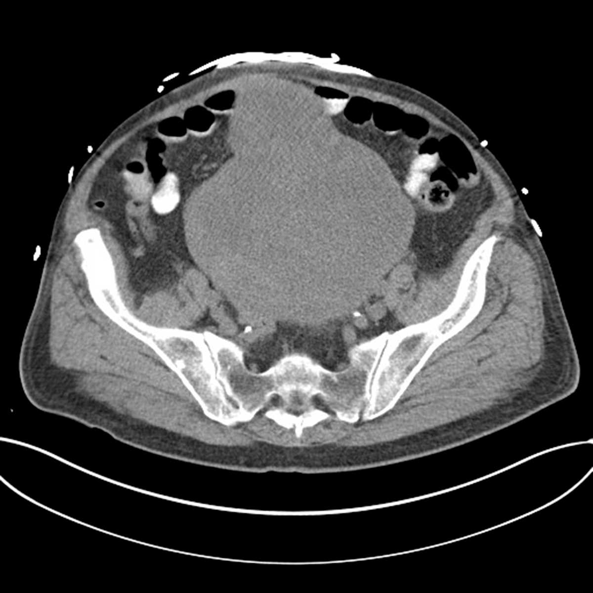
Endometrioma
Note
US images deonstrate a homogeneously hypoechoic lesion in the adnexa with low level echoes and no apperciable color flow vascularity. MRI demonstartes the lesion to be T1 hyperintense, T2 hypointense (T2 shading), and non-enhancing, in keeping with an endometrioma.
THIS IS CASE
59
OF
367


