Imaging Pearls ❯ Vascular ❯ Mycotic Aneurysms
|
-- OR -- |
|
- “The inguinal region, specifically the femoral vasculature, is a commonly used site of injection for intravenous drug users (IVDU). Repeated puncture of the vessel wall results in breakdown and subsequent arterial pseudoaneurysm- dilatations or outpouching of blood vessels, which, if left untreated, can result in fatal complications such as rupture with hemorrhage, sepsis, or even limb loss. The current modalities for arterial pseudoaneurysms include Doppler ultrasound and computed tomography (CT) angiography, both of which play important roles in management and surgical planning. However, 3D cinematic rendering (CR), a novel CT post-processing technique, offers timely, highly detailed photorealistic images that more clearly display the relation of anatomical structures, allowing for greater diagnostic confidence and precise surgical planning, particularly useful in the emergency setting.”
3D Cinematic rendering for evaluating femoral pseudoaneurysms in injection drug users
Mohammad Yasrab · Ryan C. Rizk · Linda C. Chu· Elliot K. Fishman
Emergency Radiology https://doi.org/10.1007/s10140-024-02237-x - “The inguinal region, specifically the femoral vasculature, is a The inguinal region is frequently used as a primary injection site in IVDU, therefore the femoral artery (or its branches) are the most common site where infected pseudoaneurysms develop. They can rapidly progress and often present as a surgical emergency, potentially leading to fatal consequences including rupture with hemorrhage, sepsis, and even limb-loss. Color doppler ultrasound (US) and multidetector computed tomography (CT) angiography are the most common investigations of choice for femoral pseudoaneurysms. Doppler US can swiftly confirm diagnosis by providing a dynamic view of blood flow in and out of the pseudoaneurysm sac and rule out associated venous thrombosis. On the other hand, CT imaging provides greater definition of the pseudoaneurysm and surrounding soft tissue anatomy, vasculature, and abscesses, and thus plays an important role in determining appropriate management and surgical planning.”
3D Cinematic rendering for evaluating femoral pseudoaneurysms in injection drug users
Mohammad Yasrab · Ryan C. Rizk · Linda C. Chu · Elliot K. Fishman
Emergency Radiology https://doi.org/10.1007/s10140-024-02237-x 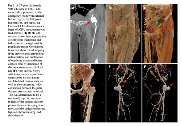
3D Cinematic rendering for evaluating femoral pseudoaneurysms in injection drug users
Mohammad Yasrab · Ryan C. Rizk · Linda C. Chu · Elliot K. Fishman
Emergency Radiology https://doi.org/10.1007/s10140-024-02237-x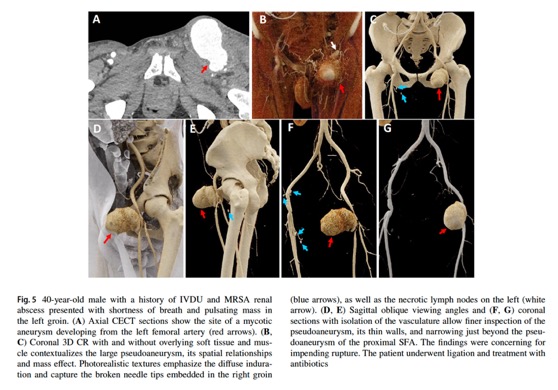
3D Cinematic rendering for evaluating femoral pseudoaneurysms in injection drug users
Mohammad Yasrab · Ryan C. Rizk · Linda C. Chu · Elliot K. Fishman
Emergency Radiology https://doi.org/10.1007/s10140-024-02237-x- “Postprocessing after CT acquisition includes the generation of select MIP, VR, and 3D CR images. 3D CR is performed at a dedicated, independent workstation that using a proprietary rendering technique developed by Siemens (syngo.via VB40 [Siemens Healthineers, Erlangen, Germany]). An NVIDIA RTX A4000 GPU powers the station. Whereas VR utilizes a local lighting model, CR employs a global illumination model, and thus Monte Carlo techniques form the basis of complex calculations and algorithms involved in determining direct and indirect paths of light rays and simulating realistic interactions with the reconstructed data. This results in a better 3D perspective, particularly at tissue interfaces. Numerous presets are available based on clinical applications, and can be further fine-tuned by various display settings, optimizing parameters, setting HU thresholds, and leveling, depending on the need of the imager and anatomic region of interest.”
3D Cinematic rendering for evaluating femoral pseudoaneurysms in injection drug users
Mohammad Yasrab · Ryan C. Rizk · Linda C. Chu · Elliot K. Fishman
Emergency Radiology https://doi.org/10.1007/s10140-024-02237-x - “By adjusting display parameters, including windowing and transparency there is greater control over which structures to emphasize or subtract. Vascular mapping with 3D CR allows for a closer inspection and characterization of the pseudoaneurysm anatomy, internal blood flow, as well as its spatial relationships with the surrounding vasculature. Evaluation of high-risk features, including subtle irregularities in texture, communication with the native vessel such as a narrow neck, as well as projections and lobulations are captured with improved depth perception in 3D, which helps ascertain the instability or imminent risk of rupture with greater confidence and thus plays a vital role in determining surgical approach and management, as alluded to in prior applications of 3D CR. For instance, a smaller pseudoaneurysm that abuts off the larger pseudoaneurysm in Fig. 6 is depicted clearly in 3D CR owing to the realistic shadowing, which wasotherwise difficult to appreciate.”
3D Cinematic rendering for evaluating femoral pseudoaneurysms in injection drug users
Mohammad Yasrab · Ryan C. Rizk · Linda C. Chu · Elliot K. Fishman
Emergency Radiology https://doi.org/10.1007/s10140-024-02237-x - “3D Cinematic Rendering (CR), an important development in CT postprocessing after traditional VR, can help thoroughly evaluate acutely developing femoral artery pseudoaneurysms in IVDU. An intuitive, photorealistic rendering of the complex spatial relationships with accurate textures, shadowing, and improved depth adds to diagnostic confidence and preoperative planning as demonstrated in the cases we present. Moving forwards, prospective studies can further evaluate its accuracy by directly comparing CR to other imaging modalities or renderingtechniques, as well as explore its utility in areas outside of diagnostic and surgical planning, such as medical education and patient counselling.”
3D Cinematic rendering for evaluating femoral pseudoaneurysms in injection drug users
Mohammad Yasrab · Ryan C. Rizk · Linda C. Chu · Elliot K. Fishman
Emergency Radiology https://doi.org/10.1007/s10140-024-02237-x


- “Brachial artery mycotic aneurysm (BAMA) is a rare condition. It can be a complication of haematogenous spread of bacterial infection. Mycotic aneurysms result from invasion and structural disruption of the arterial wall by infectious agents. Most cases described in the literature are due to intravenous drug use (IVDU) followed by bacterial endocarditis (BE).”
Mycotic Aneurysm of Brachial Artery Secondary to Infective Endocarditis.
Simson R, Jacobs T, Kulkarni SR.
EJVES Short Rep. 2019 Jul 10;46:9-11 - “BAMAs can be potentially limb or life threatening. It is important to acknowledge BE as a cause. The best therapeutic management is surgical repair after a prompt diagnosis. In this case, it was possible to perform early surgical intervention, reducing the risk of complications that could ensue from peripheral mycotic aneurysms.”
Mycotic Aneurysm of Brachial Artery Secondary to Infective Endocarditis.
Simson R, Jacobs T, Kulkarni SR.
EJVES Short Rep. 2019 Jul 10;46:9-11 - “The term “mycotic” is used to define an aneurysm/pseudoaneurysm that occurs as a consequence of infection. When applied in this context, the term is a misnomer as it used to describe all arterial infections rather than fungal infection in isolation. How-ever, it was not until the mid-19th century that mycotic pseudoaneurysms associated with other pathogens (specifically bacteria) were recognized and described. They represent a fulminant infectious process, which, if untreated, may result in systemic sepsis, rupture and exsanguination.”
Mycotic pseudoaneurysm in intravenous drug users: current insights
Richard P Stevenson et al.
Research Reports in Clinical Cardiology 2019:10 1–6 - “The bacteria associated with mycotic pseudoaneurysm in IVDU differ from those reported in historical, non-IVDU series, which is unsurprising as the pathophysiological mechanisms differ. The most commonly encountered pathogens in contemporary series in this patient population (most common), streptococci, pneumococci and enterococci. Salmonella may be encountered, but is less common. In many cases, a polymicrobial field maybe encountered, and consequently, initiation of broad-spectrum antibiotics is recommended until definitive culture results are available.”
Mycotic pseudoaneurysm in intravenous drug users: current insights
Richard P Stevenson et al.
Research Reports in Clinical Cardiology 2019:10 1–6 - ”Mycotic common femoral artery pseudoaneurysm as a consequence of inadvertent arterial self-injection in IVDU remains a common presentation in areas where there are significant populations using intravenous heroin. Diagnosis can be made on the basis of clinical grounds and is generally uncomplicated. Appropriate resuscitation with early administration of antibiotic therapy is essential. If arterial imaging is considered necessary, CT angiography is the modality of choice as it allows assessment of the retroperitoneum and allows exclusion of a significant proximal extension of sepsis. Surgical management involves drainage of sepsis and debridement of non-viable tissue.”
Mycotic pseudoaneurysm in intravenous drug users: current insights
Richard P Stevenson et al.
Research Reports in Clinical Cardiology 2019:10 1–6
- "Mycotic aneurysms are uncommon but emergent conditions in which infection of a vessel leads to a contained rupture. Progression to frank rupture, thrombosis, distal embolization, and death can occur. The widespread availability of computed tomography (CT) and its ability to obtain high-resolution, contrast-enhanced, volumetric images rapidly has made it the modality of choice for evaluating mycotic aneurysms. Three-dimensional CT visualizations can provide important information to surgeons and interventionalists prior to attempting repair of these lesions."
3D CT cinematic rendering of mycotic aneurysms Rowe SP, Chu LC, Zimmerman SL, Fishman EK
Emerg Radiol (2018). https://doi.org/10.1007/s10140-018-1643-6 - "In this case series, we demonstrate the appearance of mycotic aneurysms with the novel 3D CT visualization methodology known as cinematic rendering (CR). CR makes use of a more complex lighting model than has previously been utilized with other 3D CT techniques, allowing for enhanced surface detail and realistic shadowing effects. These features of CR may have utility in evaluating mycotic aneurysms and in pre-procedural/pre-operative planning, although a prospective study definitively evaluating this has not yet been performed."
3D CT cinematic rendering of mycotic aneurysms
Rowe SP, Chu LC, Zimmerman SL, Fishman EK
Emerg Radiol (2018). https://doi.org/10.1007/s10140-018-1643-6 - "Infected (mycotic) aneurysms are contained ruptures that occur as a result of direct infection of a vessel. These lesions can manifest in any artery in the body and can be asymptomatic, present with hemorrhage, or can produce localized or systemic symptoms of infection. While any infectious agent could, in theory, cause a mycotic aneurysm, the most common causative genera are Staphylococcus and Streptococcus."
3D CT cinematic rendering of mycotic aneurysms
Rowe SP, Chu LC, Zimmerman SL, Fishman EK
Emerg Radiol (2018). https://doi.org/10.1007/s10140-018-1643-6 - "Computed tomography (CT), particularly with an arterial phase acquisition, is the modality of choice for the evaluation of mycotic aneurysms . The acquisition of volumetric data composed of isotropic voxels with modern multidetector CT scanners facilitates the creation of 3D visualizations such as volume rendering (VR) that can assist surgeons and interventionalists in planning open surgical or endovascular repair."
3D CT cinematic rendering of mycotic aneurysms
Rowe SP, Chu LC, Zimmerman SL, Fishman EK
Emerg Radiol (2018). https://doi.org/10.1007/s10140-018-1643-6 - "CR makes use of complex path tracing of projected light rays within the context of a global illumination model in order to create images. This allows the method to take into ac- count the effect on projected rays from scatter and even from materials within adjacent voxels through which the ray does not pass. As a result, relative to traditional VR which uses ray casting and a local lighting model for image creation, CR images typically have much high levels of surface detail and realistic shadowing."
3D CT cinematic rendering of mycotic aneurysms
Rowe SP, Chu LC, Zimmerman SL, Fishman EK
Emerg Radiol (2018). https://doi.org/10.1007/s10140-018-1643-6 - "An experienced reader at our institution is able to utilize appro- priate pre-set window width and level values to create the images and interpret the findings in approximately 5 min; thus, the inclusion of CR images does not significantly impede clinical workflow."
3D CT cinematic rendering of mycotic aneurysms
Rowe SP, Chu LC, Zimmerman SL, Fishman EK
Emerg Radiol (2018). https://doi.org/10.1007/s10140-018-1643-6 - "In particular, the realistic shadowing that is intrinsic to the CR technique displays the relative relationships of vessels and other structures in an intuitive manner that is likely to prove helpful to interventionalists and surgeons."
3D CT cinematic rendering of mycotic aneurysms
Rowe SP, Chu LC, Zimmerman SL, Fishman EK
Emerg Radiol (2018). https://doi.org/10.1007/s10140-018-1643-6
- "IV drug use may result in a variety of local arterial complications at the injection site. Inadvertent arterial puncture may result in traumatic arterial dissection and even arterial occlusion with consequent acute limb ischemia. Arterial puncture may also result in formation of a false aneurysm."
Radiology of Recreational Drug Abuse
Ian G. Hagan et al.
RadioGraphics 2007; 27:919 –940 - "Cardiovascular complications include myocardial infarction, cardiomyopathy, arterial dissection, false and mycotic aneurysms, venous thromboembolic disease, and septic thrombophlebitis. Respiratory complications may involve the upper airways, lung parenchyma, pulmonary vasculature, and pleural space. ."
Radiology of Recreational Drug Abuse
Ian G. Hagan et al.
RadioGraphics 2007; 27:919 –940 - "Awareness of the imaging features of recreational drug abuse is important for the radiologist because the underlying cause may not be known at presentation and because complications affecting different body systems may coexist. Intravenous drug abuse in particular should be regarded as a multisystem disease with vascular and infective complications affecting many parts of the body, often synchronously."
Radiology of Recreational Drug Abuse
Ian G. Hagan et al.
RadioGraphics 2007; 27:919 –940 - "IV drug use may result in a variety of local arterial complications at the injection site. Inadvertent arterial puncture may result in traumatic arterial dissection and even arterial occlusion with consequent acute limb ischemia. Arterial puncture may also result in formation of a false aneurysm."
Radiology of Recreational Drug Abuse
Ian G. Hagan et al.
RadioGraphics 2007; 27:919 –940 - "Not infrequently, the nonsterile nature of injections leads to infection of false aneurysms, resulting in mycotic aneurysm formation . The presence of gas within the aneurysm is a rare but pathognomonic feature of infection and is best seen at CT."
Radiology of Recreational Drug Abuse
Ian G. Hagan et al.
RadioGraphics 2007; 27:919 –940
- “Infected aneurysms are uncommon. The aorta, peripheral arteries, cerebral arteries, and visceral arteries are involved in descending order of frequency. Staphylococcus and Streptococcus species are the most common causative pathogens. Early clinical diagnosis of infected aneurysms is challenging owing to their protean manifestations. Clinically apparent infected aneurysms are often at an advanced stage of development or are associated with complications, such as rupture. Nontreatment or delayed treatment of infected aneurysms often has a poor outcome, with high morbidity and mortality from fulminant sepsis or hemorrhage.”
Infected (Mycotic) Aneurysms: Spectrum of Imaging Appearances and Management
Lee WK et al.
November 2008 RadioGraphics 28, 1853-1868 - “Imaging features of infected aneurysms include a lobulated vascular mass, an indistinct irregular arterial wall, perianeurysmal edema, and a perianeurysmal soft-tissue mass. Perianeurysmal gas, aneurysmal thrombosis, aneurysmal wall calcification, and disrupted arterial calcification at the site of the infected aneurysm are uncommon findings.”
Infected (Mycotic) Aneurysms: Spectrum of Imaging Appearances and Management
Lee WK et al.
November 2008 RadioGraphics 28, 1853-1868 - “The prevalence of infected aortic aneurysms is 0.7%–1% of all surgically treated aortic aneurysms. The most frequently involved peripheral artery is the femoral artery, and such cases are most commonly associated with intravenous drug abuse. The prevalence of infected cerebral aneurysms is 0.7%–4% among all patients with cerebral aneurysms. The most frequently involved visceral artery is the superior mesenteric artery. Synchronous or metachronous infected aneurysms occur in 20%–36% of cases.”
Infected (Mycotic) Aneurysms: Spectrum of Imaging Appearances and Management
Lee WK et al.
November 2008 RadioGraphics 28, 1853-1868 - “Staphylococcus and Streptococcus species are the most common causes of infected aneurysms. Infected aneurysms due to methicillin-resistant Staphylococcus aureus have been reported, especially in intravenous drug abusers. Salmonella is most commonly associated with infected aortic aneurysms, especially in East Asia. Gram-negative bacteria, such as Escherichia coli, Klebsiella, and Pseudomonas, are uncommon causes of infected aneurysms that are becoming more frequent. Mycobacterium and fungi, such as Candida albicans and Aspergillus, are rare causes of infected aneurysms. Sterile blood cultures occur in 18%–50% of patients with infected aneurysms.”
Infected (Mycotic) Aneurysms: Spectrum of Imaging Appearances and Management
Lee WK et al.
November 2008 RadioGraphics 28, 1853-1868