Imaging Pearls ❯ Small Bowel ❯ Celiac Disease
|
-- OR -- |
|
- “Celiac disease is chronic intolerance of gluten that induces intestinal mucosal lesions in genetically predisposed patients. Although in most cases the symptoms and histologic abnormalities completely resolve with use of a strict gluten-free diet, com plications occur in some patients. The complications include small-bowel intussusception, ulcerative jejunoileitis, lymphoma, adenocarcinoma, hyposplenism, cavitating lymphadenopathy syndrome, and pneumatosis intestinalis.”
Celiac Disease in Adults: Evaluation with MDCT Enteroclysis
Philippe Soyer et al.
AJR 2008 191:5, 1483-1492 - “Celiac disease is now recognized as a common disease, occurring in about 1 in every 200 Americans. However, less than 10% of cases are currently diagnosed, with a diagnostic delay of more than 10 years from onset of symptoms. Celiac disease is a chronic autoimmune disorder induced in genetically susceptible individuals after ingestion of gluten proteins, which are found in wheat, rye, barley, and certain other grains. The small bowel mucosa is primarily affected, resulting in progressive degrees of villus inflammation and destruction with resulting induction of crypt hyperplasia. The destruction begins in the duodenum and over time progresses distally to the ileum. Loss of villi, which absorb fluid, and hypertrophy of crypts, which produce fluid, result in chronic fluid excess in the small bowel lumen.”
CT Findings in Adult Celiac Disease
Scholz FJ et al.
RadioGraphics 2011; 31:977–992 - “Chronic excess fluid and its effects on bowel wall structure and tone create the small bowel malabsorption pattern (MABP), which was described long ago in barium studies of patients with celiac disease. Features of the celiac disease MABP include duodenitis , dilution, dilatation, slow transit, flocculation , moulage, reversal of the jejunalileal fold pattern, and transient small bowel intussusception.”
CT Findings in Adult Celiac Disease
Scholz FJ et al.
RadioGraphics 2011; 31:977–992 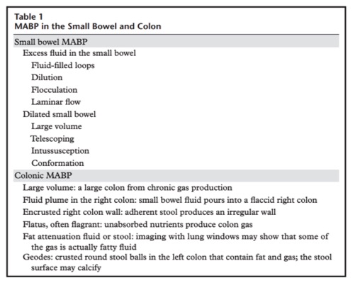
CT Findings in Adult Celiac Disease
Scholz FJ et al.
RadioGraphics 2011; 31:977–992- “Small bowel loops are often dilated and fluid filled as a result of the chronic inflammatory process. This leads to progressive dilution of enteric contrast material. Small hyperattenuating flecks of barium may be seen precipitating in the dilated small bowel loops, a phenomenon termed flocculation. The small bowel lumen contains both intrinsic physiologic fluid and administered enteric contrast material. Peristaltic waves sweeping periodically through the bowel result in variable laminar flow of these different fluid components within the flaccid bowel lumen in a recognizable pattern.”
CT Findings in Adult Celiac Disease
Scholz FJ et al.
RadioGraphics 2011; 31:977–992 - “Prominence of upper mesenteric lymph nodes is a feature of celiac disease. Autoimmune stimulation in celiac disease provokes regional lymphocytic proliferation. The duodenum and proximal jejunum are the initial organs targeted for autoimmune destruction, and nodal prominence is most marked in the upper small bowel mesentery. Mesenteric lymph node enlargement, low-attenuation lymph nodes, and cavitating lymph nodes are well-described features of celiac disease. However, cavitating or low-attenuation lymph nodes are infrequent and are found in patients with advanced symptomatic disease.”
CT Findings in Adult Celiac Disease
Scholz FJ et al.
RadioGraphics 2011; 31:977–992
- “The most common cause of villous atrophy is celiac disease. The villous atrophy results from injury to the small intestine and leads to loss of absorptive surface area, reduction of digestive enzymes, and consequential impaired absorption of micronutrients. Negative celiac serology or nonresponse to a gluten-free diet implies a broad and challenging differential diagnosis which includes Crohn's disease, enteric infections (e.g. Giardia lamblia), collagenous sprue, tropical sprue, common variable immunodeficiency, autoimmune enteropathy, hematological malignancies and medication-associated enteropathy. Regarding the latter, olmesartan medoxomil, an angiotensin receptor blocker for the management of hypertension, has been recently recognized as a cause of “sprue-like enteropathy”.”
Olmesartan-Induced Enteropathy: An Unusual Cause of Villous Atrophy Marta Eusébio et al. GE Port J Gastroenterol. 2016 Mar-Apr; 23(2): 91–95. - Legal issues with Olmesartan

- Legal issues with Olmesartan


- “Celiac disease is now recognized as a common disease, occurring in about one in every 200 Americans. However, less than 10% of cases are currently diagnosed, with a diagnostic delay of more than 10 years from onset of symptoms.”
CT Findings in Adult Celiac Disease
Scholz FJ, Afman J, Behr SC
RadioGraphics 2011; 31:977-992 - “Celiac disease is a chronic autoimmune disorder induced in genetically susceptible individuals after ingestion of gluten proteins, which are found in wheat, rye, barley, and certain other grains.”
CT Findings in Adult Celiac Disease
Scholz FJ, Afman J, Behr SC
RadioGraphics 2011; 31:977-992 - Celiac Disease: Clinical Presentation
- Abdominal pain
- Iron deficiency anemia
- Guaiac positive stools
- Diarrhea in later stages of disease
- Increased morbidity and mortality if dx is not made - Celiac Disease: CT Findings
- Small bowel loops are dilated and fluid filled
- Disease begins in duodenum and progresses from jejunum to ileum
- Fluid filled loops lead to progressive dilution of enteric contrast which is seen as precipitating flecks of contrast in dilated loops of bowel (flocculation)
- Small bowel becomes more dilated over time and this leads to intussusception especially in the pelvis
- Bowel loops especially in pelvis “conform” to each other without intervening space - Celiac Disease: CT Findings
- Nodal enlargement due to follicular hyperplasia
- Increased incidence of lymphoma because of chronic lymphatic stimulation
- Nodal enlargement best seen in upper small bowel mesentery
- Nodes may be of low CT attenuation and caveating (late finding) - Celiac Disease: CT Findings
- Colon is often dilated with excess air and poor motility
- Large volume colon due to malabsorption
- Fat fluid levels in colon due to malabsorption
- “geode” may develop in colon comprised of stool, fat and air. They may even calcify
- Spleen may atrophy over time - Cavitary Lymph Node Syndrome: Findings
- Small or dysfunctional spleen
- Low attenuation lymph nodes
- Sever villous atrophy
- May be seen in late phases of Celiac Disease