Pancreatic Mass with Calcification: Differential Dx • Serous Cystadenoma • Mucinous Cystic Neoplasm • Neuroendocrine Tumor • Solid and Pseudopapillary Neoplasm (SPEN) I• ntraductal Papillary Mucinous Neoplasm (IPMN) • Metastases to the Pancreas
Pancreatic Mass with Calcification: Differential Dx • Serous Cystadenoma • Mucinous Cystic Neoplasm • Neuroendocrine Tumor • Solid and Pseudopapillary Neoplasm (SPEN) • Intraductal Papillary Mucinous Neoplasm (IPMN) • Metastases to the Pancreas
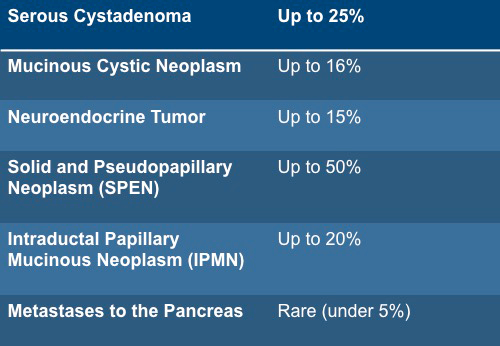
Pancreatic Mass with Calcification: Frequency
Pancreatic Mass with Calcification: Facts • Neuroendocrine tumors most commonly calcify with coarse irregular calcifications. Calcifications are usually central but can be in periphery of the mass • Serous Cystadenoma may have rim like calcification, central scar with calcification or thin calcification along radiating septae • Mucinous Cystic Neoplasm (MCN) may have peripheral calcifications or calcified septations
Pancreatic Mass with Calcification: Facts • SPEN Tumor (Solid and Pseudopapillary Neoplasm) have dense calcification more commomly in the periphery of the lesion • Intraductal Papillary Mucinous Neoplasm (IPMN) usually have peripheral or septal calcifications • Metastases to the Pancreas from Renal Cell Carcinoma may rarely calcify
Pancreatic Mass with Calcification: Pitfalls • Pancreatic adenocarcinoma that arise in an area of chronic pancreatitis may show areas of calcification due to the pancreatitis. Adenocarcinoma does not calcify. • Vascular calcifications (i.e. Splenic artery aneurysm) may simulate a neuroendocrine tumor.