Imaging Pearls ❯ Kidney ❯ Retriperitoneal Fibrosis
|
-- OR -- |
|
- “Retroperitoneal fibrosis (RPF) is a rare fibroinflammatory disease with idiopathic and secondary causes. Idiopathic disease is more common and is believed to be immune mediated; associations with autoimmune diseases and/or inflammatory disorders such as IgG4 related disease are often present. Common complications include hydronephrosis and venous stenosis and/or thrombosis. Due to its nonspecific clinical presentation, imaging is vital for diagnosis; in addition, imaging may help distinguish idiopathic from secondary causes and can aid in distinguishing early/active disease from chronic/inactive disease.”
Multimodality imaging review of retroperitoneal fibrosis.
Czerniak S, Mathur M.
Abdom Radiol (NY). 2025 Mar 4. doi: 10.1007/s00261-025-04847-6. Epub ahead of print. PMID: 4035807. - “Retroperitoneal fibrosis (RPF) is a rare disease characterized by the presence of inflammatory tissue and fibrosis centered in the retroperitoneum. The fibroinflammatory infiltrate classically develops along the anterolateral aspects of the infrarenal abdominal aorta extending inferiorly to the common iliac arteries; adjacent structures such as the ureters and other vessels are often encased resulting in hydronephrosis, venous thrombosis, and/or arterial compromise.”
Multimodality imaging review of retroperitoneal fibrosis.
Czerniak S, Mathur M.
Abdom Radiol (NY). 2025 Mar 4. doi: 10.1007/s00261-025-04847-6. Epub ahead of print. PMID: 4035807. - “Both idiopathic and secondary forms of RPF exist, with the former accounting for approximately two-thirds of cases . Idiopathic RPF is an immune-mediated process that is included as part of the chronic periaortitis disease spectrum; however, increasingly, this has been associated with autoimmune diseases and/or inflammatory disorders such as IgG4 related disease. Secondary causes of RPF include malignant disease, medications (such as ergot alkaloids), and biological agents (monoclonal antibodies such as infliximab) amongst other etiologies .”
Multimodality imaging review of retroperitoneal fibrosis.
Czerniak S, Mathur M.
Abdom Radiol (NY). 2025 Mar 4. doi: 10.1007/s00261-025-04847-6. Epub ahead of print. PMID: 4035807. - The pathogenesis of idiopathic RPF remains unclear though is likely multifactorial . The current hypothesis is that it may be a manifestation of a systemic autoimmune disease which arises as a primary aortitis; this elicits a fibroinflammatory response which subsequently extends to the adjacent retroperitoneal tissues . Inflammation in the adventitia of affected aortic segments coupled with the presence of systemic symptoms, as well as an association with autoimmune disorders and a good response to immunosuppressive therapies all support this hypothesis.
Multimodality imaging review of retroperitoneal fibrosis.
Czerniak S, Mathur M.
Abdom Radiol (NY). 2025 Mar 4. doi: 10.1007/s00261-025-04847-6. Epub ahead of print. PMID: 4035807. - “Computed tomography (CT) has become one of the preferred imaging modalities in assessing RPF, allowing a thorough evaluation of disease extent, location and morphology . In addition, CT can be used to assess for other conditions associated with RPF (IgG4 related diseases such as autoimmune pancreatitis or malignant etiologies). Even so, RPF has been reported to have no CT correlation in one third of patients with surgically proven disease . At present, the main utility of CT imaging lies in the ability to assess changes in size of the fibroinflammatory mass on serial imaging studies.”
Multimodality imaging review of retroperitoneal fibrosis.
Czerniak S, Mathur M.
Abdom Radiol (NY). 2025 Mar 4. doi: 10.1007/s00261-025-04847-6. Epub ahead of print. PMID: 4035807. - The most common imaging findings include a confluent sheet-like mass surrounding the anterior and lateral aspects of the aorta, typically centered at the aortic bifurcation with caudal extension to the common iliac arteries. Typically, there is no lateral extension beyond the lateral aspects of the psoas muscle. Cephalad extension to the level of the renal arteries or caudal extension to the sacrum may be present; additional extension to retroperitoneal organs/spaces (such as the pancreas, perinephric fat, and the retroperitoneal portions of the duodenum) has also been reported.
Multimodality imaging review of retroperitoneal fibrosis.
Czerniak S, Mathur M.
Abdom Radiol (NY). 2025 Mar 4. doi: 10.1007/s00261-025-04847-6. Epub ahead of print. PMID: 4035807. - “Radiomics is an emerging image processing technique which leverages artificial intelligence and machine learning algorithms to extract quantitative textures/features from regions of interest on radiology images. One study demonstrated that a radiomics algorithm had a discriminative accuracy of 72% in detecting residual fibrosis in retroperitoneal lymphadenopathy from testicular germ cell tumors after chemotherapy; this improved to 88% when combined with clinical predictors (prechemotherapy tumor markers, residual mass size, percentage of mass shrinkage, and the presence of teratoma elements in orchiectomy specimen.”
Multimodality imaging review of retroperitoneal fibrosis.
Czerniak S, Mathur M.
Abdom Radiol (NY). 2025 Mar 4. doi: 10.1007/s00261-025-04847-6. Epub ahead of print. PMID: 4035807. - “Confidently distinguishing between idiopathic and malignant etiologies of RPF on FDG-PET has also proved challenging. Some work on the topic, however, has shown promise; in one study which compared idiopathic RPF with lymphoma and metastatic disease to the retroperitoneum, RPF was found to have a lower frequency of high FDG uptake as well as a lower mean maximum standardized uptake value (mean SUVmax 4.8 for RPF versus 13.5 and 8.7 for lymphoma and metastases respectively). Patients with lymphoma and metastatic disease were also found to have adenopathy located at distant sites including axillary, supraclavicular, inguinal and the peritoneum.”
Multimodality imaging review of retroperitoneal fibrosis.
Czerniak S, Mathur M.
Abdom Radiol (NY). 2025 Mar 4. doi: 10.1007/s00261-025-04847-6. Epub ahead of print. PMID: 4035807. 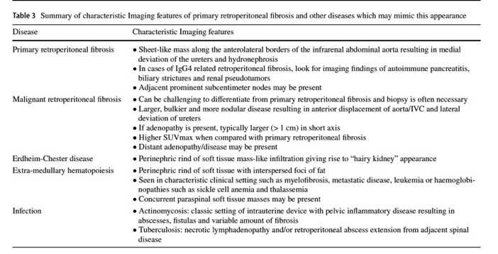
Multimodality imaging review of retroperitoneal fibrosis.
Czerniak S, Mathur M.
Abdom Radiol (NY). 2025 Mar 4. doi: 10.1007/s00261-025-04847-6. Epub ahead of print. PMID: 4035807.

- Retroperitoneal fibrosis represents fibro-inflammatory soft tissue plaque in the retroperitoneal space that often encases the aorta and one or both ureters resulting in obstruction . RPF is idiopathic in majority of the cases and has been associated with malignancy, autoimmune inflammatory disorders, GVHD, drugs, surgery, and multiple sclerosis in small number of cases . RPF usually affects males in their 4th6th decades of life and often presents with non-specific symptoms such as malaise, anorexia, and chronic backache. RPF is predominantly benign with a favorable prognosis. Small number cases, up to 8%, have been reported as malignant RPF with a poor prognosis and typical 36 months survival. Retroperitoneal fibrosis represents fibro-inflammatory soft tissue plaque in the retroperitoneal space that often encases the aorta and one or both ureters resulting in obstruction.
Imaging of ureter: a primer for the emergency radiologist
Mohd Zahid et al.
Emerg Radiol (2021). https://doi.org/10.1007/s10140-021-01930-5 - Retroperitoneal fibrosis represents fibro-inflammatory soft tissue plaque in the retroperitoneal space that often encases the aorta and one or both ureters resulting in obstruction . RPF is idiopathic in majority of the cases and has been associated with malignancy, autoimmune inflammatory disorders, GVHD, drugs, surgery, and multiple sclerosis in small number of cases . RPF usually affects males in their 4th6th decades of life and often presents with non-specific symptoms such as malaise, anorexia, and chronic backache. RPF is predominantly benign with a favorable prognosis. Small number cases, up to 8%, have been reported as malignant RPF with a poor prognosis and typical 36 months survival.
Imaging of ureter: a primer for the emergency radiologist
Mohd Zahid et al.
Emerg Radiol (2021). https://doi.org/10.1007/s10140-021-01930-5 - "CT urography reveals medial deviation and smooth narrowing of the middle one-third of one or both ureters in the lower lumbar or upper sacral region. There are variable degrees of proximal hydroureteronephrosis and delayed renal excretory function due to increased pressure. CT helps to assess the location and extent of RPF and its effect on the adjacent vascular and visceral structures. RPF may look like retroperitoneal soft tissue rind or fibrotic plaque that obliterates the periureteral and aortocaval fat planes. The important distinguishing feature of benign RPF from malignant RPF, lymphoma, and metastatic lymph nodes is that the fibrotic plaque may extend behind the aorta and anterior to the spine but rarely displaces the aorta anteriorly, although it has poor sensitivity and specificity.
Imaging of ureter: a primer for the emergency radiologist
Mohd Zahid et al.
Emerg Radiol (2021). https://doi.org/10.1007/s10140-021-01930-5 - "Degree of enhancement of benign RPF on CT correlates with fibrotic activity, avid enhancement suggests active phase, and minimal to no enhancement seen in avascular chronic plaque. Variable enhancement pattern is also seen in malignant RPF. There is significant overlap between benign RPF and malignant RPF in imaging morphology and enhancement patterns. RPF should be considered as malignant in a known abdominal primary malignancy with concomitant retroperitoneal lymph nodes. Approximately one-third surgically proven cases may have normal CT findings.
Imaging of ureter: a primer for the emergency radiologist
Mohd Zahid et al.
Emerg Radiol (2021). https://doi.org/10.1007/s10140-021-01930-5 - "Genitourinary tuberculosis is the second most common form of extrapulmonary tuberculosis and usually caused by hematogenous dissemination. It accounts for 1520% of all extrapulmonary tuberculosis cases. Genitourinary tuberculosis involves the ureter in approximately 50% of the cases. In early disease, IVU or retrograde urography shows ragged and dilated ureter and occasional filling defects of mucosal granulomas . CT urography demonstrates ureteral mural thickening with periureteral inflammatory changes. Tuberculosis tends to involve distal third of the ureter and causes multiple strictures and fibrotic changes with disease progression resulting in a characteristic beaded or corkscrew appearance . Chronic mural thickening of the ureter results in foreshortening and pipestem ureter. Tuberculosis may also present as pseudotumor due to inflammatory ureteral mural thickening and would be difficult to distinguish from malignancy on imaging . In a small number of cases, ureteral calcifications can be seen.
Imaging of ureter: a primer for the emergency radiologist
Mohd Zahid et al.
Emerg Radiol (2021). https://doi.org/10.1007/s10140-021-01930-5
- Extramedullary Hematopoeisis: Common Causes
- myelofibrosis
- diffuse osseous metastatic disease replacing the bone marrow
- leukaemia
- sickle cell disease
- thalassemia. - Infiltration of the Perinephric Space: Differential Diagnosis
- Retroperitoneal fibrosis
- Erdheim-Chester disease
- Lymphoma
- Extramedullary hematopoiesis
- Liposarcoma
- Metastases (melanoma is classic)
- Hematoma (often post trauma or biopsy)