Imaging Pearls ❯ Kidney ❯ Erdheim-Chester Disease
|
-- OR -- |
|
- “Erdheim-Chester disease (ECD) is a rare multiorgan histiocytosis with diverse clinical manifestations. Initially described as “lipoid granulomatosis” by William Chester and Jakob Erdheim in 1930 , it was named after its founders by Henry Jaffe in 1972. ECD was previously considered inflammatory histiocytosis, but the discovery of activating mutations in the mitogen-activated protein kinase (MAPK) signaling pathway has established ECD as a histiocytic neoplasm, although inflammation contributes to organ damage . Hence, ECD was included in the “histiocytic and dendritic cell neoplasms” category in the 2016 World Health Organization classification of hematopoietic tumors. Similarly, ECD along with Langerhans cell histiocytosis (LCH) has been included in the Langerhans group in the 2016 revised Histiocyte Society classification of histiocytosis, as both entities share mutations in the MAPK pathway.”
Imaging in Erdheim-Chester Disease
Yashant Aswani, et al.
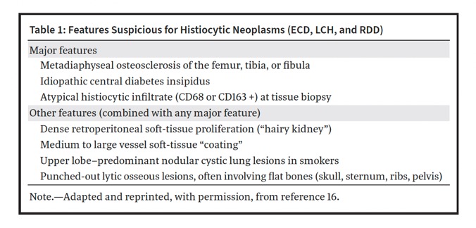
RadioGraphics 2024; 44(9):e240011 - - The pattern of bilateral symmetric metadiaphyseal regions of osteosclerosis, especially around the knees, is virtually pathognomonic for ECD. These regions reveal intense uptake at scintigraphy and FDG PET/ CT, again a finding nearly exclusive to ECD.
- ECD in the retroperitoneum manifests as infiltrative soft-tissue masses in bilateral perirenal (hairy kidneys) and posterior pararenal spaces.
- Myocardial infiltration has a predilection for the right atrium and atrioventricular sulcus and manifests as a focal mural mass (pseudotumor).
- Chest radiographic findings may be normal. Pulmonary involvement is, however, seen at CT in 50% of patients and often shows smooth interlobular septal thickening. The histiocytic infiltrate is perilymphatic in distribution and causes smooth thickening of the interlobular septa, peribronchovascular interstitium, and visceral pleura.
- Intracranial lesions in ECD, including intra-axial, meningeal, perivascular, and pituitary infundibulum lesions, are rarely isolated, and patients who harbor these lesions almost always have osteosclerosis of the paranasal sinus walls and/or orbital disease.
Imaging in Erdheim-Chester Disease
Yashant Aswani, et al.
RadioGraphics 2024; 44(9):e240011 
Imaging in Erdheim-Chester Disease
Yashant Aswani, et al.
RadioGraphics 2024; 44(9):e240011- “Retroperitoneal and renal involvement in ECD is seen in 50%–60% of patients. Although patients with renal involvement are frequently asymptomatic, they may present with abdominal pain and dysuria . ECD in the retroperitoneum manifests as infiltrative soft-tissue masses in bilateral perirenal (hairy kidneys) and posterior pararenal spaces. On CT images, this soft tissue is isoattenuating relative to skeletal muscle and shows mild postcontrast enhancement. At MRI, it is iso- to hypointense relative to skeletal muscle on T1- and T2-weighted images and shows mild postcontrast enhancement. Because the normal renal parenchyma shows intense uptake of FDG, the sensitivity of FDG PET/CT for detection of retroperitoneal involvement, in particular perinephric ECD, is not as high as contrast-enhanced CT.”
Imaging in Erdheim-Chester Disease
Yashant Aswani, et al.
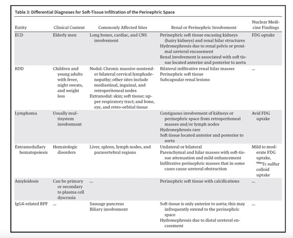
RadioGraphics 2024; 44(9):e240011 - “Perinephric soft-tissue infiltrates, other than ECD, can also be seen in lymphoma, retroperitoneal fibrosis (RPF), RDD, amyloid, and extramedullary hematopoeisis (EMH) . ECD and lymphoma may involve the perinephric and retroaortic spaces and show uptake at FDG PET/ CT. Contiguous involvement of the kidneys or perinephric space from retroperitoneal masses or lymph nodes is a common pattern of renal lymphoma. Contrarily, ECD rarely affects the lymph nodes.”
Imaging in Erdheim-Chester Disease
Yashant Aswani, et al.
RadioGraphics 2024; 44(9):e240011 - “Although ECD and RPF cause hydronephrosis, the former is epicentered around the renal hila and proximal ureters, whereas the latter is epicentered around aortic bifurcation and hence affects the distal ureters. As RPF extends more superiorly, it lies anterior and lateral to the aorta and rarely infiltrates the renal hila and perinephric spaces. Immunoglobulin G4 (IgG4)–related RPF may also show pancreatic involvement. Renal involvement in RDD most commonly manifests as bilateral hilar masses.”
Imaging in Erdheim-Chester Disease
Yashant Aswani, et al.
RadioGraphics 2024; 44(9):e240011 
Imaging in Erdheim-Chester Disease
Yashant Aswani, et al.
RadioGraphics 2024; 44(9):e240011- “ECD is a multiorgan inflammatory histiocytic neoplasm that most frequently affects the skeletal system and produces virtually pathognomonic osteosclerosis around the knees that shows increased radiotracer uptake on nuclear medicine studies. Other typical imaging findings comprise perinephric infiltrates (hairy kidneys), periaortic soft tissue (coated aorta), and a right atrial pseudotumor. However, radiologic findings are interpreted in conjunction with clinical and histologic features to establish the diagnosis. In addition to aiding in diagnosis, imaging also helps to determine the extent of disease, target sites for tissue sampling, assess treatment response,and predict the prognosis.”
Imaging in Erdheim-Chester Disease
Yashant Aswani, et al.
RadioGraphics 2024; 44(9):e240011
- Erdheim–Chester disease: Renal Findings
- enlarged kidneys
- perirenal infiltrations shown as characteristic “hairy kidney” (70%)
- Renal sinus involvement
- Thickening of the ureters
- Soft tissue thickening of the renal arteries - “In summary, urinary involvement in ECD was mostly bilateral and symmetric. Early identification of the perirenal hairy kidney sign is critical for accurate diagnosis and timely treatment. Understanding of the CT findings and progression as well as correct diagnosis based on radiological findings, have significant implications for guiding ECD treatment.”
Urinary involvement in Erdheim–Chester disease: computed tomography imaging findings
Zhe Wu et al.
Abdominal Radiology (2021) 46:4324–4331 - Erdheim Chester Disease: Renal Changes
- Perirenal fat effaced or infiltrated by soft tissue
- Usually bilateral and symmetric
- Infiltration of para-aortic regions is also common - Erdheim Chester Disease: Vascular Changes
- Periaortic infiltration which is usually circumferential and nonocclusive
- May involve aorta from root thru iliac vessels
- May involve the renal arteries - Erdheim Chester Disease: Skeletal Changes
- Bilateral symmetric osteosclerosis of metaphyses and diaphyses especially in long bone.


