Imaging Pearls ❯ Esophagus ❯ Perforation
|
-- OR -- |
|
- “Atrioesophageal fistula (AEF) after atrial fibrillation ablation is the most serious and feared complication. AEF is difficult to diagnose, and delays in diagnosis are common. Highly variable symptoms usually do not start to appear for 1 week or longer postprocedure, and when they appear, the patient often presents to a community hospital staffed by providers with little knowledge of AEF . Postablation esophageal perforation can present with variations including true AEF, pericardioesophageal fistula, and mediastinal-esophageal fistula. True AEF usually has a precursor esophageal lesion, which is often neglected or confused with pericarditis, a more common complication associated with atrial fibrillation ablation. Esophageal lesions eventually ulcerate and may progress into a direct connection between esophagus and left atrium, causing air embolism with stroke, sepsis, and sometimes esophageal bleeding. AEF is rare but is often fatal, especially if not treated, with mortality rates ranging from 40% to 100%.”
Recognition, Management, and Prevention of Atrioesophageal Fistula
Catanzaro JN, Assis FR, Verma A, Tandri H, Tilz RR, Spragg DD, Calkins H, Fishman EK, Deneke T..
JACC Clin Electrophysiol. 2024 Apr 13:S2405-500X(24)00165-8. doi: 10.1016/j.jacep.2024.02.022. Epub ahead of print. PMID: 38703161. 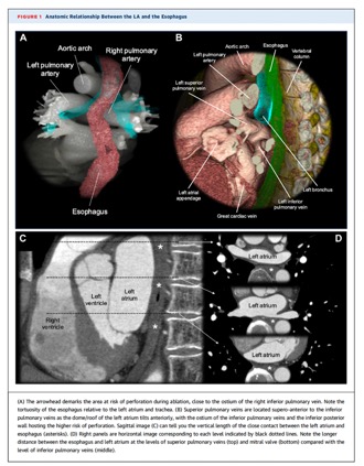
Recognition, Management, and Prevention of Atrioesophageal Fistula
Catanzaro JN, Assis FR, Verma A, Tandri H, Tilz RR, Spragg DD, Calkins H, Fishman EK, Deneke T..
JACC Clin Electrophysiol. 2024 Apr 13:S2405-500X(24)00165-8. doi: 10.1016/j.jacep.2024.02.022. Epub ahead of print. PMID: 38703161.

- Complications of Esophageal Rupture and Fistula Formation
● Aortoesophageal fistula
● Tracheoesophageal fistula
● Chronic paraesophageal abscess as a result of remote esophageal perforation
● Thoracic discitis/osteomyelitis secondary to esophageal perforation
● Duplication cyst rupture into distal esophagus - Aortoesophageal Fistula
● Rare and often fatal cause of upper gastrointestinal bleeding
● Most commonly due to localized rupture of thoracic aortic aneurysm or secondary complication of aneurysm repair or graft infection
○ Less common causes include foreign body ingestion, esophageal malignancy and syphilis
● Classic clinical triad first described by Chiari:
○ Dysphagia or midthoracic pain radiating to back
○ Sentinel hemorrhage followed by asymptomatic interval
○ Massive upper gastrointestinal hemorrhage leading to exsanguination
● High clinical suspicion is imperative for prompt detection with MDCT, triage and successful surgical repair
● MDCT is sensitive for the detection of fistula formation
○ Presence of focal esophageal wall thickening, extraluminal air, contrast extravasation, perianeurysmal hematoma or pseudoaneurysm should raise concern for aortoenteric fistula - Tracheoesophageal Fistula
● Majority of esophagorespiratory fistulas in adults are acquired
○ Direct invasion by intrathoracic malignancies, mainly esophageal carcinoma, account for greater than 60% of cases
■ Reported in up to 5-10% of patients with advanced esophageal cancer; increased risk if history of prior radiation
○ Other causes include prolonged intubation, foreign body ingestion, esophageal instrumentation, trauma and granulomatous infection
● Suspect esophagorespiratory fistula in patients with known esophageal cancer and recurrent pneumonia
● Esophagogram is usually definitive and can differentiate aspiration versus fistula
○ MDCT is useful if fluoroscopy is equivocal and may define malignancy or fistulous tract
● Prognosis is poor with treatment often being palliative
○ Endobronchial stent placement, esophageal bypass via gastrostomy/jejunostomy, or surgical bypass or correction
- “The development of an esophageal-airway fistula is a life-threatening complication of esophageal cancer or secondary to esophageal trauma, infections, or radiochemotherapy. Initial symptoms most often include cough, aspiration and fever, frequently culminating in pneumonia. More than half such fistulas involve the trachea; alternatively, a connection with the left or right main or lower lobe bronchus may be formed. Generally patients with esophageal airway fistulas are treated with covered stents to seal off the leak. CT may be necessary to localize the fistula and to aid in treatment planning. CT can also be used to detect pleuro-plumonary or mediastinal inflammatory reactions to esophageal fistulae .”
Dedicated multi-detector CT of the esophagus: spectrum of diseases Ahmed Ba-Ssalamah et al. Abdominal Imaging Jan 2009, Volume 34, Issue 1, pp 3–18 - “Esophageal injuries include penetrating injuries, blunt traumatic perforation, iatrogenic perforation as well as spontaneous perforation due to a sudden rise in intraluminal pressure during vomiting (so-called Boerhaave syndrome). Most often, esophageal perforation occurs during endoscopic investigation of malignant disease and presents a difficult problem. Esophageal diseases, such as strictures, achalasia, and tumors predispose the esophagus to perforation.”
Dedicated multi-detector CT of the esophagus: spectrum of diseases Ahmed Ba-Ssalamah et al. Abdominal Imaging Jan 2009, Volume 34, Issue 1, pp 3–18
- “Gastroesophageal reflux disease is a common cause of noncardiac chest pain. Esophagitis related to the ingestion of caustic substances, irradiation, medication, or infection also may result in acute chest pain. In severe esophagitis, full-thickness esophageal necrosis may lead to perforation with associated complications. Contrast material–enhanced esophagography and endoscopy remain the reference standards for the evaluation of esophagitis. CT may be performed when the diagnosis is unclear or when a complication is suspected. Whatever the cause of severe esophagitis, its CT appearance is predominantly characterized by diffuse esophageal thickening, submucosal edema, and mucosal enhancement.”
CT Features of Esophageal Emergencies Catherine A. Young, MD et al RadioGraphics 2008 28:6, 1541-1553 - “Transmural perforation may occur in a variety of settings. Its clinical presentation and CT appearance are similarly variable, depending on the mechanism of injury, the site and size of perforation, and the time elapsed since the onset of symptoms . Iatrogenic perforation of the esophagus is increasingly common, with therapeutic endoscopic procedures such as stricture dilation and stent placement being the leading causes. Perforation also occurs, albeit infrequently, as a complication of surgical procedures such as gastric fundoplication, esophageal myotomy, thyroidectomy, and anterior cervical diskectomy ”
CT Features of Esophageal Emergencies Catherine A. Young, MD et al RadioGraphics 2008 28:6, 1541-1553 - “Transmural perforation may occur in a variety of settings. Its clinical presentation and CT appearance are similarly variable, depending on the mechanism of injury, the site and size of perforation, and the time elapsed since the onset of symptoms. Latrogenic perforation of the esophagus is increasingly common, with therapeutic endoscopic procedures such as stricture dilation and stent placement being the leading causes . Perforation also occurs, albeit infrequently, as a complication of surgical procedures such as gastric fundoplication, esophageal myotomy, thyroidectomy, and anterior cervical diskectomy.”
CT Features of Esophageal Emergencies Catherine A. Young, MD et al RadioGraphics 2008 28:6, 1541-1553 - “CT is a useful adjunct to barium esophagography and direct visualization in the diagnosis and evaluation of esophageal emergencies. CT evaluation of the esophagus requires a high index of suspicion in appropriate clinical settings and attention to findings that may be subtle yet significant. Given the orientation of the esophagus, multiplanar reformatted images are ideally suited to this task and often yield a better appreciation of the extent of disease and its relation to adjacent structures. An awareness of the CT findings associated with the spectrum of acute esophageal disease will promote the radiologist’s ability to accurately diagnose esophageal emergencies, thereby reducing delays in diagnosis that are likely to have a negative effect on outcomes.”
CT Features of Esophageal Emergencies Catherine A. Young, MD et al RadioGraphics 2008 28:6, 1541-1553