Imaging Pearls ❯ Colon ❯ Intussusception
|
-- OR -- |
|
- “Intussusception is defined as the invagination of one segment of the bowel into an immediately adjacent segment of the bowel. Idiopathic ileocolic intussusception is the most common form in children and is typically managed with nonoperative reduction via pneumatic and/or hydrostatic enemas. In the adult population, intussusception is uncommon and occurs more often in the small intestine than in the colon. It is associated with lead point pathology in most symptomatic cases presenting as bowel obstruction. When lead point pathology is present in adult small bowel intussusception, it is usually benign, though when malignant it is most frequently due to diffuse metastatic disease, for example, melanoma. In contrast, adult ileocolic and colonic intussusception lead point pathology is most frequently primary adenocarcinoma when malignant.”
Intestinal Intussusception: Etiology, Diagnosis, and Treatment.
Marsicovetere P, Ivatury SJ, White B, Holubar SD.
Clin Colon Rectal Surg. 2017 Feb;30(1):30-39. - “The mean age of intussusception in adults is 50 years with no gender predominance. In direct contrast to pediatric etiologies, adult intussusception is associated with an identifiable cause in 90% of symptomatic cases with an idiopathic cause in 10% of cases.Benign or malignant neoplasms cause two-thirds of cases with a lead point; the remaining cases are caused by infections, postoperative adhesions, Crohn granulomas, intestinal ulcers (Yersinia), and congenital abnormalities such as Meckel diverticulum. Of the cases caused by neoplasms, 50% of them are malignant. Independent predictors of malignancy include colonic intussusception and anemia.”
Intestinal Intussusception: Etiology, Diagnosis, and Treatment.
Marsicovetere P, Ivatury SJ, White B, Holubar SD.
Clin Colon Rectal Surg. 2017 Feb;30(1):30-39. - “Most adult intussusceptions arise from the small bowel, and most lesions are benign with a rate of 50 to 75% in most series. The most common lesions are Meckel diverticulum and adhesions. Benign tumors include lymphoid hyperplasia, lipomas, leiomyomas, hemangiomas, and polyps. Other conditions that predispose to small bowel intussusception include anorexia, nervosa, and malabsorption syndromes, as increased flaccidity of the bowel wall facilitates invagination. Supratherapeutic anticoagulation therapy may cause submucosal hemorrhages that can lead to intussusception. Less commonly, malignant tumors may act as lead points with metastatic disease (i.e., carcinomatosis) being the most common. In several reports, approximately 50% of malignant lesions causing small bowel intussusception were metastatic (miliary) melanomas.Malignant intraluminal causes of small bowel intussusception include primary leiomyosarcomas, adenocarcinoma, GIST tumors, carcinoid tumors, neuroendocrine tumors, and lymphomas.”
Intestinal Intussusception: Etiology, Diagnosis, and Treatment.
Marsicovetere P, Ivatury SJ, White B, Holubar SD.
Clin Colon Rectal Surg. 2017 Feb;30(1):30-39. - “Adult intussusception less commonly occurs in the colon than in the small bowel and accounts for only 20 to 25% of all intussusceptions in most reported case series. The most common malignant cause of colonic intussusception is primary colonic adenocarcinoma and the most common benign cause is colonic lipoma. Contrary to the small intestine, several reports indicate that colonic intussusception is more likely to have a malignant lead point due to the increased prevalence of malignancies in the colon versus the small bowel. However, other studies conflict with this and suggest the proportion of benign and malignant lesions causing colonic intussusception is similar to that of small bowel intussusception. Ileocolic intussusception in adults is a unique variant in which nearly 100% of cases have a malignant lead point, namely, cecal adenocarcinoma involving the ileocecal valve.”
Intestinal Intussusception: Etiology, Diagnosis, and Treatment.
Marsicovetere P, Ivatury SJ, White B, Holubar SD.
Clin Colon Rectal Surg. 2017 Feb;30(1):30-39. - “In adults, the clinical presentation of intussusception can be nonspecific, rarely presenting with the classic triad of abdominal pain, palpable mass, and bloody stool. Instead, it presents with symptoms of small or large bowel obstruction. The most common presenting symptom is abdominal pain, with associated symptoms consistent with partial obstruction: nausea, vomiting, obstipation, gastrointestinal bleeding, change in bowel habits, constipation, or bloating. Wang et al found abdominal cramping pain in nearly 80% of patients as a leading symptom; a palpable abdominal mass, however, was found in less than 9%.”
Intestinal Intussusception: Etiology, Diagnosis, and Treatment.
Marsicovetere P, Ivatury SJ, White B, Holubar SD.
Clin Colon Rectal Surg. 2017 Feb;30(1):30-39. 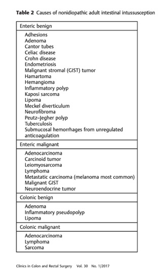
- “In adults, the clinical presentation of intussusception can be nonspecific, rarely presenting with the classic triad of abdominal pain, palpable mass, and bloody stool. Instead, it presents with symptoms of small or large bowel obstruction. The most common presenting symptom is abdominal pain, with associated symptoms consistent with partial obstruction: nausea, vomiting, obstipation, gastrointestinal bleeding, change in bowel habits, constipation, or bloating. Wang et al found abdominal cramping pain in nearly 80% of patients as a leading symptom; a palpable abdominal mass, however, was found in less than 9%.”
Colonic Intussusception: Clinical and Radiographic Features
Gollub MJ
AJR 2011; 196:W580–W585 - Colonic intussusception is caused by a malignancy more frequently than is smallbowel intussusception, because of the greater prevalence of malignant tumors in the colon (e.g., primary adenocarcinoma, lymphoma, and metastatic disease to the colon) than in the small bowel. Benign colonic lesions constitute about 30% of colonic intussusceptions and include lipoma, benign stromal tumors, adenomatous polyps, endometriosis, and previous anastomoses. Ileocecal intussusception can occur from a lead point in the colon, ileum, or appendix. Causes may include lipoma, inflammatory fibroid polyp, hamartomatous polyp, lymphoma, adenocarcinoma of the ileum or cecum, or even Meckel diverticulum. Inflammatory appendiceal conditions can also cause intussusception, but the most commonly reported entity is a mucocele.
Colonic Intussusception: Clinical and Radiographic Features
Gollub MJ
AJR 2011; 196:W580–W585 - “Among my patients, only one with colonic intussusception ultimately presented emergently with severe cramping abdominal pain, vomiting, and diarrhea. Most cases required surgery but not usually emergently. Other malignant intussusceptions spontaneously resolved after chemotherapy. As in other series, most cases at my institution occurred in the proximal colon, either right or transverse. Possible explanations include a greater tendency in the proximal colon for polypoid neoplasms and a longer colonic mesentery, which allows greater colonic mobility. Obstruction was a rare feature in this series."
Colonic Intussusception: Clinical and Radiographic Features
Gollub MJ
AJR 2011; 196:W580–W585 - “Intussusception is the invagination of a bowel loop with its mesenteric fold (intussusceptum) into the lumen of a contiguous portion of bowel (intussuscipiens) as a result of peristalsis. Intraluminal polypoid lesions have a greater tendency to cause invagination of the bowel as peristalsis drags the lesion forward. Although the exact mechanism precipitating intussusception, especially intussusception without a lead point, is not well understood, this condition has been ascribed to dysrhythmic contractions.”
Adult Intestinal Intussusception: CT Appearances and Identification of a Causative Lead Point
Young H. Kim, et al.
RadioGraphics 2006; 26:733–744 - Adenocarcinoma of the colon is the most common malignant neoplasm associated with colonic intussusception .Typical signs and symptoms of adenocarcinoma of the colon include bleeding, obstruction, a palpable abdominal mass, and abdominal pain. The individual layers of the intussuscepted bowel wall are more easily distinguished from the lead mass in this intussusception. Differentiation of the lead mass from bowel wall edema at CT is generally easier in large bowel intussusception than in small bowel intussusception due to the greater caliber of the colon.
Adult Intestinal Intussusception: CT Appearances and Identification of a Causative Lead Point
Young H. Kim, et al.
RadioGraphics 2006; 26:733–744 - “Lipomas are the most common benign cause of colocolic intussusception in adults. Next to adenomatous polyps, these mesenchymal tumors are the most common benign tumors of the colon. Lipomas of the colon are within the submucosa in 90% of cases, are usually solitary, and may be sessile or pedunculated . Lipomas are often discovered incidentally at endoscopic or radiologic examination and can easily be diagnosed with CT due to their typical fat attenuation. Close observation of consecutive axial images can help avoid misinterpreting entrapped mesentery and subserosal fat as a lipoma. Multiplanar reformation may be used to confirm the diagnosis of intussusception when axial views raise suspicion for such a diagnosis. Lipomas are almost always asymptomatic until they cause abdominal pain, sometimes due to intussusception.”
Adult Intestinal Intussusception: CT Appearances and Identification of a Causative Lead Point
Young H. Kim, et al.
RadioGraphics 2006; 26:733–744 - Causes of Adult Intestinal Lead Point Intussusception (Colon)
Benign
- Lipoma
- adenomatous polyp
Malignant
- Adenocarcinoma
- lymphoma,
- metastasis
Idiopathic
- Postoperative adhesion,
- motility disorder

- Ileocecal Intussusception : Key Facts
- In children can be due to nodes or lymphoid hyperplasia or mass
- Meckels is rare cause but can occur as do benign and malignant tumors