Imaging Pearls ❯ Colon ❯ Acute Abdomen
|
-- OR -- |
|
- "Peritonitis is the most common clinical manifestation of abdominal TB. Infection of the peritoneum is usually secondary to hematogenous spread from a pulmonary focus, from adjacent organs such as the intestine or the fallopian tube or ruptured necrotic lymph node. Liver cirrhosis, human immunodeficiency virus–positive, and chronic renal failure patients on continuous ambulatory peritoneal dialysis are at an increased risk for peritoneal TB."
Imaging Spectrum of Extra thoracic Tuberculosis
Rout, Abhijit A. et al.
Radiologic Clinics , Volume 54 , Issue 3 , 475 - 501 - "Based on the pattern of ascites, omental and peritoneal tubercles, and associated inflammatory and fibrotic response, TB peritonitis has been traditionally classified into 3 categories."
Imaging Spectrum of Extrathoracic Tuberculosis
Raut, Abhijit A. et al.
Radiologic Clinics , Volume 54 , Issue 3 , 475 - 501 - "Wet ascitic type-This is the most common variety of peritoneal TB seen among 90% of the cases with significant free or loculated high-density ascitic fluid on CT scan.".
Imaging Spectrum of Extrathoracic Tuberculosis
Raut, Abhijit A. et al.
Radiologic Clinics , Volume 54 , Issue 3 , 475 - 501 - "Fibrotic fixed type-This relatively less frequent variety of peritoneal TB is characterized by mesenteric and omental thickening, tuberculous deposits, and matted bowel loops.".
Imaging Spectrum of Extrathoracic Tuberculosis
Raut, Abhijit A. et al.
Radiologic Clinics , Volume 54 , Issue 3 , 475 - 501 - "Dry or 'plastic' type-his is a rare variety showing peritoneal nodules, fibrous peritoneal reaction, and dense adhesions. Ultrasonography can accurately demonstrate small quantities of loculated or free ascitic fluid. Multiple thin, complete or incomplete septae can be seen with echogenic debris being frequent in the loculated ascites. However, these features are can also be seen in malignancy, chronic infective peritonitis, and hemoperitoneum. CT scan shows loculated or free, high attenuation ascitic fluid (20–45 Hounsfield unites [HU]), omental thickening/nodularity, and thickened inflamed mesentery associated with mesenteric adenopathy."
Imaging Spectrum of Extrathoracic Tuberculosis
Raut, Abhijit A. et al.
Radiologic Clinics , Volume 54 , Issue 3 , 475 - 501 - "The common routes of spread of tubercle bacilli to the gastrointestinal tract (GIT) include hematogeneous spread from the primary lung lesion, ingestion of infected sputum from active pulmonary focus, direct spread from adjacent organs, or through lymphatic spread from infected lymph nodes. Although TB can involve any part of the GIT, the most common target site of involvement is the IC region. This is likely owing to several factors, including relative stasis, abundant lymphoid tissue, and closer contact of the bacilli with the mucosa in this region."
Imaging Spectrum of Extrathoracic Tuberculosis
Raut, Abhijit A. et al.
Radiologic Clinics , Volume 54 , Issue 3 , 475 - 501 - "The IC region is the most frequent site for bowel TB. The frequency of bowel involvement decreases both proximally and distally from the IC region. Multiple strictures, adhesions, and bowel obstruction are the most the most common complications. Perforation followed by fistulae formation and intestinal bleeding may be seen but are uncommon."
Imaging Spectrum of Extrathoracic Tuberculosis
Raut, Abhijit A. et al.
Radiologic Clinics , Volume 54 , Issue 3 , 475 - 501 - "CT scan is the modality of choice for the evaluation of abdominal TB. Mural thickening of the IC region is frequent in GIT TB. It may be limited to the terminal ileum or caecum, or it may involve both .The thickening may be symmetric or asymmetric. Asymmetric thickening of the IC valve and medial wall of the caecum may have an exophytic extension and this may engulf the terminal ileum. Fat stranding and inflammation is seen in the pericecal region and adjacent mesentery. There may be associated bowel obstruction and/or perforation."
Imaging Spectrum of Extrathoracic Tuberculosis
Raut, Abhijit A. et al.
Radiologic Clinics , Volume 54 , Issue 3 , 475 - 501 - "Hepatosplenic TB occurs secondary to hematogeneous spread of disease from active tuberculous focus elsewhere in the body. Commonly, it presents as miliary form in association with miliary pulmonary TB. Miliary TB manifests as hepatomegaly with multiple tiny low-attenuation foci without significant postcontrast enhancement on CT scan. The macronodular form of hepatosplenic TB is uncommon and manifests as a few hypodense lesions with ill-defined margins."
Imaging Spectrum of Extrathoracic Tuberculosis
Raut, Abhijit A. et al.
Radiologic Clinics , Volume 54 , Issue 3 , 475 - 501

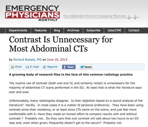
- The routine use of contrast (both oral and IV, and certainly rectal) is unnecessary for the majority of abdominal CT scans performed in the ED. At least that is what the literature says over and over. Unfortunately, many radiologists disagree. Is their objection based on a sound analysis of the literature? Hardly. In most cases it is a matter of personal preference. They have been using contrast since their residency, or at least since CTs came on the scene, and just feel more comfortable with it. Have they made an honest effort to compare results with and without contrast ? Probably not. Do they care that oral contrast will add about two hours to an ED stay and, even when given, frequently doesn’t get to the cecum? Probably not.
- “In the care of elderly patients, CT is accurate for diagnosing the cause of acute abdominal pain, particularly when it is of surgical origin, regardless of the availability of clinical and biologic findings. Thus CT interpretation should not be delayed until complete clinicobiologic data are available, and the images should be quickly transmitted to the emergency physician so that appropriate therapy can be begun.”
Acute abdominal pain in elderly patients: effect of radiologist awareness of clinicobiologic information on CT accuracy
Millet I et al.
AJR Am J Roentgenol. 2013 Dec;201(6):1171-8 - “In both the entire cohort (87.4% vs. 85.3%, p = 0.07) and the surgical group (94% vs. 91%, p = 0.15), there was no significant difference in CT accuracy between diagnoses made when the radiologist was aware and those made when the radiologist was not aware of the clinicobiologic findings. Agreement between the CT diagnosis and the final diagnosis was excellent whether or not the radiologist was aware of the clinicobiologic findings.”
Acute abdominal pain in elderly patients: effect of radiologist awareness of clinicobiologic information on CT accuracy
Millet I et al.
AJR Am J Roentgenol. 2013 Dec;201(6):1171-8 - “The cases of 333 consecutively registered patients 75 years old or older presenting to the emergency department with acute abdominal pain and who underwent CT were retrospectively reviewed by two radiologists blinded or not to the patient's clinicobiologic results. Diagnostic accuracy was calculated according to the level of correctly classified cases in both the entire cohort and a surgical subgroup and was compared between readings performed with and without knowledge of the clinicobiologic findings. Agreement between each reading and the reference diagnosis and interobserver agreement were assessed with kappa statistics.”
Acute abdominal pain in elderly patients: effect of radiologist awareness of clinicobiologic information on CT accuracy
Millet I et al.
AJR Am J Roentgenol. 2013 Dec;201(6):1171-8 - “Axial and coronal reformations of 64-section multidetector row CT have equal sensitivity and specificity for the diagnosis of acute abdominal pathology. However, coronal reformations improved the diagnostic confidence for all readers but most significantly for the least experienced. Therefore, radiology departments with residents should consider routinely generating coronal images in patients with acute abdominal pain.”
Acute abdomen: Added diagnostic value of coronal reformations with 64-slice multidetector row computed tomography.
Zangos S et al.
Acad Radiol. 2007 Jan;14(1):19-27. - “For the most inexperienced reader, the coronal reformations were helpful in 95% of cases, while for the most experienced reader, the coronal reformations were helpful in 35% of the cases. The coronal images were deemed helpful in an average of 62.3% of the cases for the four readers. However, diagnosing subtle pathology in the abdominal wall was difficult on coronal reformations alone. Overall, coronal reformations improved diagnostic confidence and interobserver agreement over axial images alone for visualization of normal abdominal structures and in the diagnosis of abdominal pathology.”
Acute abdomen: Added diagnostic value of coronal reformations with 64-slice multidetector row computed tomography.
Zangos S et al.
Acad Radiol. 2007 Jan;14(1):19-27.

