Imaging Pearls ❯ Chest ❯ Screening CT
|
-- OR -- |
|
- ”We analyzed GI cancer deaths among participants in New York State (1992–2010), exploring demographics and GI cancer distribution. Radiologists retrospectively reviewed pancreatic cancer cases within 24 months post-LDCT, comparing findings with original reports. Among 10,150 participants, 189 died from GI cancers; mean age 75, mostly male smokers. Pancreatic cancer (41.8%) led, followed by esophageal (17.5%) and colon cancer (16.9%). Median time between baseline LDCT and death was 116 months (9.7 years). 82/189 (43.4%) participants died within 5 years of their last LDCT screening, with pancreatic cancer again prominent (45.1%). In 79 pancreatic cancer deaths, 17.7% occurred within 24 months post-LDCT. A re-review identified previously undetected pancreatic findings, with 4 out of 14 participants (28.6%) showing abnormalities. This underscores the potential of lung cancer screening programs to provide insights beyond lung health. This study of over 10,000 participants in a lung cancer screening program reveals that they are at risk for GI cancer deaths, particularly pancreatic cancer. Re-reviews of LDCT scans revealed previously undocumented pancreatic findings in a third of participants who died from pancreatic cancer, underscoring the need to identify, document, and follow up on these findings. ”
GI cancer mortality in participants in low dose CT screening for lung cancer with a focus on pancreatic cancer.
Gros L, Yip R, Zhu Y, Li P, Paksashvili N, Sun Q, Yankelevitz DF, Henschke CI.
Sci Rep. 2024 Dec 2;14(1):29851. doi: 10.1038/s41598-024-76322-z. PMID: 39617764; 
GI cancer mortality in participants in low dose CT screening for lung cancer with a focus on pancreatic cancer.
Gros L, Yip R, Zhu Y, Li P, Paksashvili N, Sun Q, Yankelevitz DF, Henschke CI.
Sci Rep. 2024 Dec 2;14(1):29851. doi: 10.1038/s41598-024-76322-z. PMID: 39617764;- “It is crucial to carefully examine, document, and monitor the pancreas during lung cancer screenings. This study underscores the potential of lung cancer screening programs to provide valuable insights beyond lung health. Incorporating technological advancements, such as AI tools, could significantly enhance the effectiveness of these screenings and pave the way for including pancreatic cancer detection in lung cancer protocols, providing a comprehensive health check.”
GI cancer mortality in participants in low dose CT screening for lung cancer with a focus on pancreatic cancer.
Gros L, Yip R, Zhu Y, Li P, Paksashvili N, Sun Q, Yankelevitz DF, Henschke CI.
Sci Rep. 2024 Dec 2;14(1):29851. doi: 10.1038/s41598-024-76322-z. PMID: 39617764; - “Of the 82 participants who died from GI cancer within 5 years of their last LDCT, 17 (20.7%) had lesions that, upon retrospective re-review could have been considered as signs of cancer (median time before deaths: 27.8 months). However, it remains uncertain whether these findings, and their thorough exploration, could have influenced the course of events as over half of the cases had suspicious findings of already advanced disease.”
GI cancer mortality in participants in low dose CT screening for lung cancer with a focus on pancreatic cancer.
Gros L, Yip R, Zhu Y, Li P, Paksashvili N, Sun Q, Yankelevitz DF, Henschke CI.
Sci Rep. 2024 Dec 2;14(1):29851. doi: 10.1038/s41598-024-76322-z. PMID: 39617764; - Of the 79 participants who died of pancreatic cancer, two had pancreatic lesions identified on the original report of their last LDCT screening. These included a cystic structure in the tail of the pancreas (discovered 49 months before death), and a dilatation of the main pancreatic duct (noted 58 months before death). Both of these signs are indicators that should have raised suspicion of pancreatic cancer in the 2 participants26,27. The other 77 (97.5%) participants who died of pancreatic cancer had no reported pancreatic lesions.
GI cancer mortality in participants in low dose CT screening for lung cancer with a focus on pancreatic cancer.
Gros L, Yip R, Zhu Y, Li P, Paksashvili N, Sun Q, Yankelevitz DF, Henschke CI.
Sci Rep. 2024 Dec 2;14(1):29851. doi: 10.1038/s41598-024-76322-z. PMID: 39617764
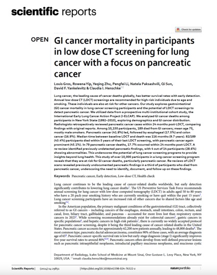
- Among 10,150 participants, 189 died from GI cancers; mean age 75, mostly male smokers. Pancreatic cancer (41.8%) led, followed by esophageal (17.5%) and colon cancer (16.9%). Median time between baseline LDCT and death was 116 months (9.7 years). 82/189 (43.4%) participants died within 5 years of their last LDCT screening, with pancreatic cancer again prominent (45.1%). In 79 pancreatic cancer deaths, 17.7% occurred within 24 months post-LDCT. A re-review identified previously undetected pancreatic findings, with 4 out of 14 participants (28.6%) showing abnormalities. This underscores the potential of lung cancer screening programs to provide insights beyond lung health. This study of over 10,000 participants in a lung cancer screening program reveals that they are at risk for GI cancer deaths, particularly pancreatic cancer. Re-reviews of LDCT scans revealed previously undocumented pancreatic findings in a third of participants who died from pancreatic cancer, underscoring the need to identify, document, and follow up on these findings.
GI cancer mortality in participants in low dose CT screening for lung cancer with a focus on pancreatic cancer
Louis Gros, Rowena Yip, Yeqing Zhu, et al.
Scientific Reports | (2024) 14:29851 - A re-review identified previously undetected pancreatic findings, with 4 out of 14 participants (28.6%) showing abnormalities. This underscores the potential of lung cancer screening programs to provide insights beyond lung health. This study of over 10,000 participants in a lung cancer screening program reveals that they are at risk for GI cancer deaths, particularly pancreatic cancer. Re-reviews of LDCT scans revealed previously undocumented pancreatic findings in a third of participants who died from pancreatic cancer, underscoring the need to identify, document, and follow up on these findings.
GI cancer mortality in participants in low dose CT screening for lung cancer with a focus on pancreatic cancer
Louis Gros, Rowena Yip, Yeqing Zhu, et al.
Scientific Reports | (2024) 14:29851 - “On re-review of the baseline LDCTs for these 14 participants (median time 31.5 months, IQR 20–75, before death), 4 participants (28.6%) (compared to 0/14 in the original report, p = 0.098) were found to have pancreatic lesions: one with calcifications, two with atrophy, and one with both calcifications and atrophy. Among them, 3/4 had stable pancreatic findings on the last LDCT (median 26 months later, IQR 14-37.5), while one showed worsening atrophy.”
GI cancer mortality in participants in low dose CT screening for lung cancer with a focus on pancreatic cancer
Louis Gros, Rowena Yip, Yeqing Zhu, et al.
Scientific Reports | (2024) 14:29851 - “This underscores the need to follow a well-defined protocol for detecting and interpreting pancreatic findings, along with providing appropriate follow-up recommendations, especially for participants with a significant smoking history and diabetes; associated with an increased risk of pancreatic cancer41. The retrospective nature of this study, despite the prospective data collection, presents an inherent limitation. Relying on mortality data may introduce a bias, especially for pancreatic cancer, which, while relatively uncommon, is exceptionally deadly. This bias could lead to a distortion in the apparent frequency of pancreatic cancer compared to other cancers within our cohort. The entire pancreas may not be fully visualized in LDCT scans performed for lung cancer screening. Furthermore, image quality and soft tissue resolution may be affected by the use of LDCT. However, our study demonstrates that pancreatic findings can still be detected using LDCT. ”
GI cancer mortality in participant in low dose CT screening for lung cancer with a focus on pancreatic cancer
Louis Gros, Rowena Yip, Yeqing Zhu, et al.
Scientific Reports | (2024) 14:29851
- RESULTS: Of screening tests with a SIF detected, 12 228 (89.1%) had a SIF considered reportable to the RC, with a higher proportion of reportable SIFs among those with a positive screen result for lung cancer (7632 [94.1%]) compared with those with a negative screen result (4596 [81.8%]). The most common SIFs reported included emphysema (8677 [43.0%] of 20 156 SIFs reported), coronary artery calcium (2432 [12.1%]), and masses or suspicious lesions (1493 [7.4%]). Masses included kidney (647 [3.2%]), liver (420 [2.1%]), adrenal (265 [1.3%]), and breast (161 [0.8%]) abnormalities.
CONCLUSIONS AND RELEVANCE This case series study found that SIFs were commonly reported in the LDCT arm of the National Lung Screening Trial, and most of these SIFs were considered reportable to the RC and likely to require follow-up. Future screening trials should standardize SIF reporting.
Significant Incidental Findings in the National Lung Screening Trial
Ilana F. Gareen et al
JAMA Intern Med. doi:10.1001/jamainternmed.2023.1116 - Question What were the types of significant incidental findings (SIFs) detected on low-dose computed tomography screening examinations in the National Lung Screening Trial?
Findings In this case series study of 26 455 participants who underwent screening with low-dose computed tomography in the National Lung Screening Trial, 8954 (33.8%) had a SIF reported. Most screening tests with a SIF (12 228 [89.1%]) had at least 1 abnormality considered reportable to the referring clinician; the most common SIFs included emphysema (8677 of 20 156 [43.0%]), coronary artery calcium (2432 [12.1%]), and masses (1493 [7.4%]).
Meaning The results of this case series study suggest that SIFs should be reported in a consistent manner and that SIF management should follow established guidelines to potentially minimize costs to patients, clinicians, and the health care system.
Significant Incidental Findings in the National Lung Screening Trial
Ilana F. Gareen et al
JAMA Intern Med. doi:10.1001/jamainternmed.2023.1116 - “In this case series study, slightly more than one-third of all NLST LDCT screening participants had a SIF detected. Most of these SIFs were classified as reportable to the RC for additional evaluation. This large number of potentially clinically important SIFs may be associated with a heavy burden of follow-up care and testing, with implications for patients, clinicians, and the health care system. At the same time, the discovery of these SIFs can potentially present an opportunity for early detection of non– lung cancer conditions in a high-risk population.”
Significant Incidental Findings in the National Lung Screening Trial
Ilana F. Gareen et al
JAMA Intern Med. doi:10.1001/jamainternmed.2023.1116
- “Lung cancer is the second most common cancer and the leading cause of cancer death in the US. In 2020, an estimated 228 820 persons were diagnosed with lung cancer, and 135 720 persons died of the disease. The most important risk factor for lung cancer is smoking. Increasing age is also a risk factor for lung cancer. Lung cancer has a generally poor prognosis, with an overall 5-year survival rate of 20.5%. However, early-stage lung cancer has a better prognosis and is more amenable to treatment.”
Screening for Lung Cancer: US Preventive Services Task Force Recommendation Statement.
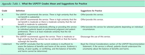
US Preventive Services Task Force. JAMA. 2021;325(10):962–970. - The USPSTF recommends annual screening for lung cancer with LDCT in adults aged 50 to 80 years who have a 20 pack-year smoking history and currently smoke or have quit within the past 15 years. Screening should be discontinued once a person has not smoked for 15 years or develops a health problem that substantially limits life expectancy or the ability or willingness to have curative lung surgery. (B recommendation) This recommendation replaces the 2013 USPSTF statement that recommended annual screening for lung cancer with LDCT in adults aged 55 to 80 years who have a 30 pack-year smoking history and currently smoke or have quit within the past 15 years.
Screening for Lung Cancer: US Preventive Services Task Force Recommendation Statement.
US Preventive Services Task Force. JAMA. 2021;325(10):962–970. - Smoking and older age are the 2 most important risk factors for lung cancer.The risk of lung cancer in persons who smoke increases with cumulative quantity and duration of smoking and with age but decreases with increasing time since quitting for persons who formerly smoked.The USPSTF considers adults aged 50 to 80 years who have a 20 pack-year smoking history and currently smoke or have quit within the past 15 years to be at high risk and recommends screening for lung cancer with annual LDCT in this population.
Screening for Lung Cancer: US Preventive Services Task Force Recommendation Statement.
US Preventive Services Task Force. JAMA. 2021;325(10):962–970. - Shared decision-making is important when clinicians and patients discuss screening for lung cancer. The benefit of screening varies with risk because persons at higher risk are more likely to benefit. Screening does not prevent most lung cancer deaths; thus, smoking cessation remains essential. Lung cancer screening has the potential to cause harm, including false-positive results and incidental findings that can lead to subsequent testing and treatment, including the anxiety of living with a lung lesion that may be cancer. Overdiagnosis of lung cancer and the risks of radiation exposure are harms, although their exact magnitude is uncertain. The decision to undertake screening should involve a thorough discussion of the potential benefits, limitations, and harms of screening.
Screening for Lung Cancer: US Preventive Services Task Force Recommendation Statement.
US Preventive Services Task Force. JAMA. 2021;325(10):962–970. - This recommendation replaces the 2013 USPSTF recommendation on screening for lung cancer. In 2013 the USPSTF recommended annual screening for lung cancer with LDCT in adults aged 55 to 80 years who have a 30 pack-year smoking history and currently smoke or have quit within the past 15 years (abbreviated as A-55-80-30-15).For this updated recommendation, the USPSTF has changed the age range and pack-year eligibility criteria and recommends annual screening for lung cancer with LDCT in adults aged 50 to 80 years who have a 20 pack-year smoking history and currently smoke or have quit within the past 15 years (A-50-80-20-15).
Screening for Lung Cancer: US Preventive Services Task Force Recommendation Statement.
US Preventive Services Task Force. JAMA. 2021;325(10):962–970.
- Lung Cancer Screening
• Can CT be used for the early detection of lung cancer?
• Should CT be used for the early detection of lung cancer?
• What is the current status of CT for the early detection of lung cancwr? - Who should be screened?
• Adults aged 55-80 years of age
• 30 pack year history of smoking
• Currently smoke or have quit within the past 15 years
• Exclusions
- Patients who stopped smoking over 15 years ago
- Patient with comorbidities that make the patient a non-surgical candidate - Lung Cancer CT Screening Protocols
• 64 slice MDCT or better
• Low dose scan protocols with 100 kV and 30-100 mA depending on the scanner and availability of iterative reconstruction software
• Slice thickness of 1-1.5 mm slice thickness
• Post processing with MPR (especially coronal) and MIP imagings - Lung Cancer Screening: Challenges
Extra Parenchymal Findings
• Cardiovascular
• Pulmonary (not a nodule)
• Adrenal nodules
• Hepatic lesions
• Renal lesions - “Clinically significant incidental findings on LDCT scans for lung cancer screening are common and their potential impact should be included in the shared decision making process. Screening programs should develop a standard approach for the evaluation of these findings, and consider the financial impact when seeking infrastructure support for screening program implementation.”
The Frequency of Incidental Findings and Subsequent Evaluation in Low-Dose CT Scans for Lung Cancer Screening. Morgan L et al Ann Am Thorac Soc. 2017 Apr 19. doi: 10.1513 (in press) - “The most commonly reported incidental findings were pulmonary (69.6%), cardiovascular (67.5%) and gastrointestinal (25.9%). Fifteen percent of the scans had an incidental finding that resulted in further evaluation. The majority of patients who underwent further testing had cardiovascular findings (10.3%), less frequently thyroid or adrenal nodules (2.1%), hepatic lesions (0.9%), renal mass (0.6%), or pulmonary disease (0.6%). The most frequently ordered investigations were echocardiography (n=9), cardiac stress test (n=9) and CT angiography (n=6). Reimbursement for the screening process, evaluation and treatment of screen detected findings, averaged $817 per screened patient.”
The Frequency of Incidental Findings and Subsequent Evaluation in Low-Dose CT Scans for Lung Cancer Screening. Morgan L et al Ann Am Thorac Soc. 2017 Apr 19. doi: 10.1513 (in press) - “The National Lung Cancer Screening Trial (NLST) is the largest RCT (n=53,454) and compared three annual LDCT scans (the first of which is termed the baseline LDCT) with three annual posterior- anterior CXRs. The NLST trial was conducted at 33 sites in the United States, with almost 75% of sites considered tertiary care hospitals.”
Screening for lung cancer Sateia HF, Choi Y, Stewart RW, Peairs KS Semin Oncol. 2017 Feb;44(1):74-82. - “The incidence of lung cancer in the LDCT group was 645 cases per 100,000 person-years compared with lung cancer incidence in the CXR group of 572 cases per 100,000 person- years (rate ratio, 1.13; 95% CI, 1.03–1.23). The relative reduc- tion in lung cancer mortality with LDCT screening was 20.0% (95% CI, 6.8– 26.7; P 1⁄4 .004) over 5.4 years of follow up, with 247 lung cancer deaths per 100,000 person-years in the LDCT group and 309 deaths per 100,000 person-years in the CXR group.”
Screening for lung cancer Sateia HF, Choi Y, Stewart RW, Peairs KS Semin Oncol. 2017 Feb;44(1):74-82. - “The number needed to screen (NNS) to prevent one lung cancer death was 320. All-cause mortality was also decreased by 6.7% with a NNS of 219 to prevent one death.”
Screening for lung cancer Sateia HF, Choi Y, Stewart RW, Peairs KS Semin Oncol. 2017 Feb;44(1):74-82. - Limitations and Challenges of Screening
(1) the work up that ensues, one that may include invasive procedures that are potentially harmful especially in the case of a false positive result
(2) unnecessary treatment
(3) the anxiety it generates
(4) radiation exposure
(5) less inclination to quit
(6) cost - Lung Cancer Screening Guidelines
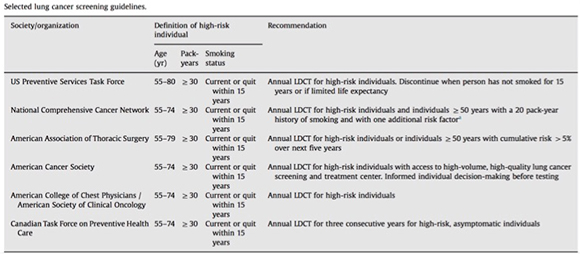
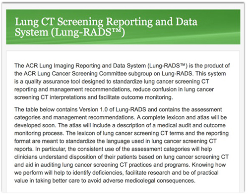
- “The ACR has developed a set of tools necessary for radiologists to take the lead on the front lines of lung cancer screening. The ACR Lung Cancer Screening Center designation is built upon the ACR CT accreditation program and requires use of Lung-RADS or a similar structured reporting and management system. This designation provides patients and referring providers with the assurance that they will receive high-quality screening with appropriate follow-up care.”
ACR CT Accreditation Program and the Lung Cancer Screening Program Designation Ella A. Kazerooni et al. JACR Vol 12, Issue 1, January 2015, Pages 38–42 - ACR Lung Cancer Screening Center designation: additional requirements
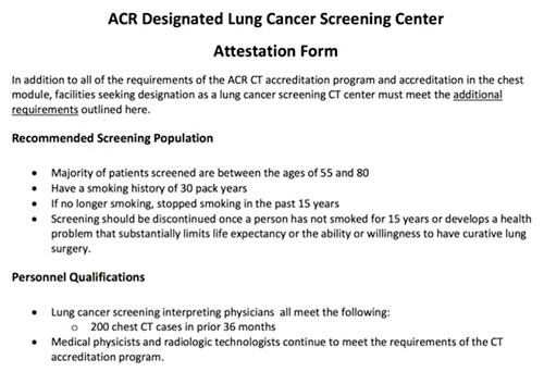
• Definition of eligible and appropriate screening population
• Incorporation of smoking cessation
• Physician qualification of at least 200 chest CT exams in the prior 36 months
• Structured reporting and management tool, such as Lung-RADS
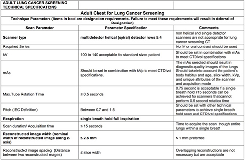
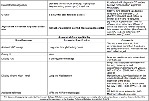
• Multidetector, helical (spiral) scanner; low-dose CT protocol must have a CTDIvol of ≤3 mGy for a standard-size patient (5’7”; 154 lb)
• Exposure techniques must be adjusted for patient size
• Participation in the ACR Dose Index Registry is recommended
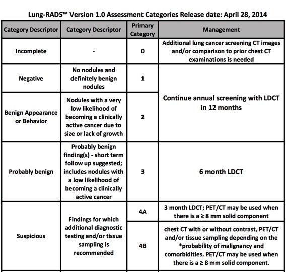
• Use and submit a low-dose CT protocol that meets the criteria outlined in the ACR- Society of Thoracic Radiology Practice Parameter for the Performance and Reporting of Lung Cancer Screening Thoracic CT - PURPOSE: The aim of this study was to assess the effect of applying ACR Lung-RADS in a clinical CT lung screening program on the frequency of positive and false-negative findings.
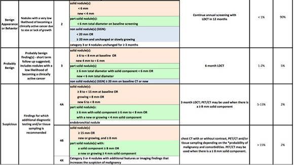
CONCLUSIONS: The application of ACR Lung-RADS increased the positive predictive value in our CT lung screening cohort by a factor of 2.5, to 17.3%, without increasing the number of examinations with false-negative results. Performance of ACR Lung-RADS in a Clinical
CT Lung Screening Program McKee BJ et al J Am Coll Radiol. 2016 Feb;13(2 Suppl):R25-9. - “ACR Lung-RADS reduced the overall positive rate from 27.6% to 10.6%. No false negatives were present in the 152 patients with >12-month follow-up reclassified as benign. Applying ACR Lung-RADS increased the positive predictive value for diagnosed malignancy in 1,603 patients with follow-up from 6.9% to 17.3%.”
Performance of ACR Lung-RADS in a Clinical CT Lung Screening Program McKee BJ et al J Am Coll Radiol. 2016 Feb;13(2 Suppl):R25-9. 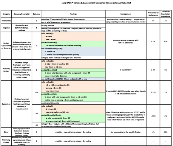
- Lung Cancer Screening: The Downside
- False positive exams
- Overdiagnosis
- Complications of diagnostic evaluation
- Radiation exposure - Lung Cancer Screening: The Downside
False positive exams-Based on solid evidence, at least 98% of all positive low-dose helical computed tomography screening exams (but not all) do not result in a lung cancer diagnosis. False-positive exams may result in unnecessary invasive diagnostic procedures. - Lung Cancer Screening: The Downside
Overdiagnosis- Based on solid evidence, a modest but non-negligible percentage of lung cancers detected by screening chest x-ray and/or sputum cytology appear to represent overdiagnosed cancer; the magnitude of overdiagnosis appears to be between 5% and 25%. - Lung Cancer Screening: The Downside
Complications of diagnostic evaluation-These cancers result in unnecessary diagnostic procedures and also lead to unnecessary treatment. Harms of diagnostic procedures and treatment occur most frequently among long-term and/or heavy smokers because of smoking-associated comorbidities that increase risk propagation. - Lung Cancer Screening: The Downside
Radiation Exposure- exams even though low dose require radiation exposure and no dose is the same as low dose - NLST Trial: Facts
The NLST included 33 centers across the United States. Eligible participants were between the ages of 55 years and 74 years at randomization, had a history of at least 30 pack-years of cigarette smoking, and, if former smokers, had quit within the past 15 years. A total of 53,454 persons were enrolled; 26,722 persons were randomly assigned to receive screening with LDCT, and 26,732 persons were randomly assigned to receive screening with chest x-ray.
Lung Cancer Screening (PDQ®) from National Cancer Institute at the NIH - NLST Trial: Facts
Any noncalcified nodule found on LDCT measuring at least 4 mm in any diameter and any noncalcified nodule or mass identified on x-ray images were classified as positive, although radiologists had the option of calling a final screen negative if a noncalcified nodule had been stable on the three screening exams. The LDCT group had a substantially higher rate of positive screening tests than did the radiography group (round 1, 27.3% vs. 9.2%; round 2, 27.9% vs. 6.2%; and round 3, 16.8% vs. 5.0%). - NLST Trial: Facts
Overall, 39.1% of participants in the LDCT group and 16.0% in the radiography group had at least one positive screening result. Of those who screened positive, the false-positive rate was 96.4% in the LDCT group and 94.5% in the chest radiography group. This was consistent across all three rounds. - NLST Trial: Facts
In the LDCT group, 649 cancers were diagnosed after a positive screening test, 44 after a negative screening test, and 367 among participants who either missed the screening or received the diagnosis after the completion of the screening phase. In the radiography group, 279 cancers were diagnosed after a positive screening test, 137 after a negative screening test, and 525 among participants who either missed the screening or received the diagnosis after the completion of the screening phase. Three hundred fifty-six deaths from lung cancer occurred in the LDCT group, and 443 deaths from lung cancer occurred in the chest x-ray group, with a relative reduction in the rate of death from lung cancer of 20% (95% confidence interval) with LDCT screening. Overall, mortality was reduced by 6.7% (95% CI). The number needed to screen with LDCT to prevent one death from lung cancer was 320. - NLST Trial: Facts
In the LDCT group, 649 cancers were diagnosed after a positive screening test, 44 after a negative screening test, and 367 among participants who either missed the screening or received the diagnosis after the completion of the screening phase. In the radiography group, 279 cancers were diagnosed after a positive screening test, 137 after a negative screening test, and 525 among participants who either missed the screening or received the diagnosis after the completion of the screening phase. - NLST Trial: Facts
Three hundred fifty-six deaths from lung cancer occurred in the LDCT group, and 443 deaths from lung cancer occurred in the chest x-ray group, with a relative reduction in the rate of death from lung cancer of 20% (95% confidence interval) with LDCT screening. Overall, mortality was reduced by 6.7% (95% CI). The number needed to screen with LDCT to prevent one death from lung cancer was 320. - NLST Trial: Facts
The number needed to screen with LDCT to prevent one death from lung cancer was 320. 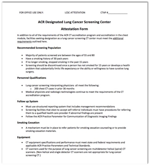
- Lung Cancer At Presentation
- “We estimated that screening for lung cancer with low-dose CT would cost $81,000 per QALY (quality-adjusted life-years) gained, but we also determined that modest changes in our assumptions would greatly alter this figure. The determination of whether screening outside the trial will be cost-effective will depend on how screening is implemented.”
Cost-effectiveness of CT screening in the National Lung Screening Trial.
Black WC et al.
N Engl J Med. 2014 Nov 6;371(19):1793-802 - Questions about Lung Cancer Screening?
What is it?
How do we do it?
Should we do it and if yes, why should we do it?
Is it cost effective?
Is it going to be available soon? - “As compared with no screening, screening with low-dose CT cost an additional $1,631 per person (95% confidence interval [CI], 1,557 to 1,709) and provided an additional 0.0316 life-years per person (95% CI, 0.0154 to 0.0478) and 0.0201 QALYs per person (95% CI, 0.0088 to 0.0314). The corresponding ICERs were $52,000 per life-year gained (95% CI, 34,000 to 106,000) and $81,000 per QALY gained (95% CI, 52,000 to 186,000). However, the ICERs varied widely in subgroup and sensitivity analyses.”
Cost-effectiveness of CT screening in the National Lung Screening Trial.
Black WC et al.
N Engl J Med. 2014 Nov 6;371(19):1793-802 - BACKGROUND:
The National Lung Screening Trial (NLST) showed that screening with low-dose computed tomography (CT) as compared with chest radiography reduced lung-cancer mortality. We examined the cost-effectiveness of screening with low-dose CT in the NLST.
CONCLUSIONS:
We estimated that screening for lung cancer with low-dose CT would cost $81,000 per QALY gained, but we also determined that modest changes in our assumptions would greatly alter this figure. The determination of whether screening outside the trial will be cost-effective will depend on how screening is implemented. - BACKGROUND: The National Lung Screening Trial (NLST) showed that screening with low-dose computed tomography (CT) as compared with chest radiography reduced lung-cancer mortality. We examined the cost-effectiveness of screening with low-dose CT in the NLST.
CONCLUSIONS: We estimated that screening for lung cancer with low-dose CT would cost $81,000 per QALY gained, but we also determined that modest changes in our assumptions would greatly alter this figure. The determination of whether screening outside the trial will be cost-effective will depend on how screening is implemented.
Cost-effectiveness of CT screening in the National Lung Screening Trial.
Black WC et al.
N Engl J Med. 2014 Nov 6;371(19):1793-802 - Can we improve the results of the NLST by adjusting guidelines?
Change lung nodule size criteria
Change clinical requirements for participation in the study
change follow up recommendations
change use of non-imaging resources - Potential drawbacks to lung cancer screening
Detect cancers (including BAC) that may be indolent
Detect nodules which are benign but require follow up or even surgery - “Computed tomography (CT) screening for lung cancer has been associated with a high frequency of false positive results because of the high prevalence of indeterminate but usually benign small pulmonary nodules. The acceptability of reducing false-positive rates and diagnostic evaluations by increasing the nodule size threshold for a positive screen depends on the projected balance between benefits and risks.”
Projected outcomes using different nodule sizes to define a positive CT lung cancer screening examination.
Gierada DS et al.
J Natl Cancer Inst. 2014 Oct 18;106(11). - “Raising the nodule size threshold for a positive screen would substantially reduce false-positive CT screenings and medical resource utilization with a variable impact on screening outcomes.”
Projected outcomes using different nodule sizes to define a positive CT lung cancer screening examination.
Gierada DS et al.
J Natl Cancer Inst. 2014 Oct 18;106(11). - “In 64% of positive screens (11598/18141), the largest nodule was 7 mm or less in greatest transverse diameter. By increasing the threshold, the percentages of lung cancer diagnoses that would have been missed or delayed and false positives that would have been avoided progressively increased, for example from 1.0% and 15.8% at a 5 mm threshold to 10.5% and 65.8% at an 8 mm threshold, respectively. The projected reductions in postscreening follow-up CT scans and invasive procedures also increased as the threshold was raised. Differences across nodules sizes for lung cancer histology and stage distribution were small but statistically significant. There were no differences across nodule sizes in survival or mortality.”
Projected outcomes using different nodule sizes to define a positive CT lung cancer screening examination.
Gierada DS et al.
J Natl Cancer Inst. 2014 Oct 18;106(11). - “More than 18% of all lung cancers detected by LDCT in the NLST seem to be indolent, and overdiagnosis should be considered when describing the risks of LDCT screening for lung cancer.”
Overdiagnosis in low-dose computed tomography screening for lung cancer.
Patz EF Jr et al.
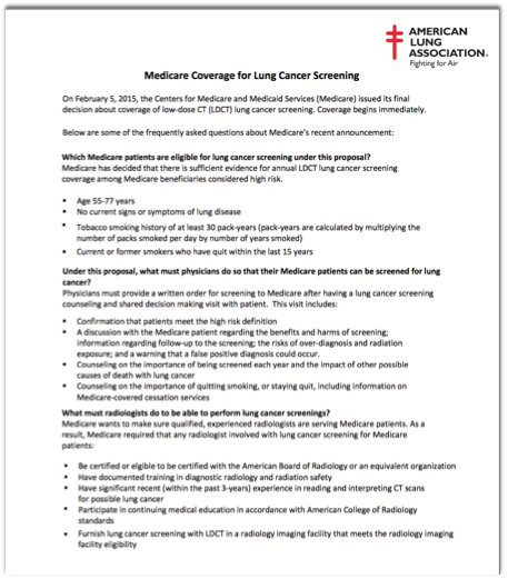
JAMA Intern Med. 2014 Feb 1;174(2):269-74 - Who should be screened?
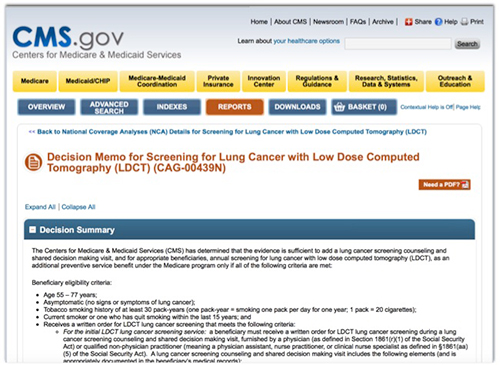
Age 55-77 years
No current signs or symptoms of lung disease
Tobacco smoking history of at least 30 pack-years (pack-years are calculated by multiplying the number of packs smoked per day by number of years smoked)
Current or former smokers who have quit within the last 15 years - What must the referring doctor do to get a screening Lung CT?
Physicians must provide a written order for screening to Medicare after having a lung cancer screening counseling and shared decision making visit with patient. This visit includes: 4 steps
- Confirmation that patients meet the high risk definition
- A discussion with the Medicare patient regarding the benefits and harms of screening; information regarding follow-up to the screening; the risks of over-diagnosis and radiation exposure; and a warning that a false positive diagnosis could occur.
- Counseling on the importance of being screened each year and the impact of other possible causes of death with lung cancer
- Counseling on the importance of quitting smoking, or staying quit, including information on Medicare-covered cessation services - What are the requirement for radiology/radiologists?
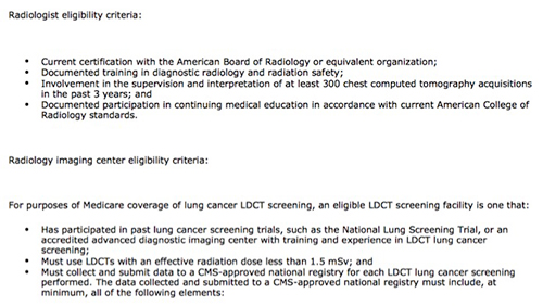
Be certified or elgible to be certified with the American Board of Radiology or an equivalent organization
Have documented training in diagnostic radiology and radiation safety
Have significant recent (within the past 3-years) experience in reading and interpreting CT scans for possible lung cancer - What are the requirement for radiology/radiologists?
Participate in continuing medical education in accordance with American College of Radiology standards
Furnish lung cancer screening with LDCT in a radiology imaging facility that meets the radiology imaging facility eligibility 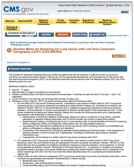
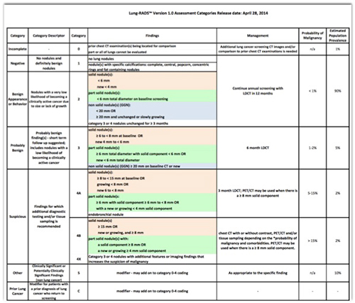
- Benign or Probably Benign

- Suspicious Nodule

- PURPOSE: The present study was performed to evaluate the utility of axial, coronal and sagittal multiplanar reformations (MPR) and maximum intensity projections (MIP) in the detection of pulmonary nodules as compared to axial standard reconstructions (SR).
CONCLUSION: MIP reformations on the basis of MDCT data sets are superior in the depiction and diagnosis of pulmonary nodules as compared to axial standard reconstructions and MPR.
Multidetector-row CT of the lungs: Multiplanar reconstructions and maximum intensity projections for the detection of pulmonary nodules
Eibel R et al.
Rofo. 2001 Sep;173(9):815-21. - “The NLST has demonstrated that the screening of high-risk populations with the use of LDCT reduces lung cancer mortality. Reporting LDCT may appear unproblematic but radiologists involved in LCS should be aware that the interpretation of low-dose CT scans is not as easy as it may initially seem. Several considerations such as different nodule detection, tools and measurement assessments should be taken into account in order to avoid false-negative diagnoses as well as unnecessary follow-up examinations. Moreover, knowledge of the significance of different nodule types is essential, as different nodules require different approaches for their detection, measurement and management.“
Low-dose CT: technique, reading methods and image interpretation
Cristiano Rampinelli et al.
Cancer Imaging. 2012; 12(3): 548–556. - “The NLST has demonstrated that the screening of high-risk populations with the use of LDCT reduces lung cancer mortality. Reporting LDCT may appear unproblematic but radiologists involved in LCS should be aware that the interpretation of low-dose CT scans is not as easy as it may initially seem. Several considerations such as different nodule detection, tools and measurement assessments should be taken into account in order to avoid false-negative diagnoses as well as unnecessary follow-up examinations.”
Low-dose CT: technique, reading methods and image interpretation
Cristiano Rampinelli et al.
Cancer Imaging. 2012; 12(3): 548–556. - “The final institution interpretations agreed with the center in 895 of the 1000 interpretations. Compared with the center, the frequency of positive results was higher at eight of the 10 institutions. The most frequent reason of discrepant interpretations was not following the protocol (n = 55) and the least frequent was not identifying a nodule (n = 3).”
CT screening for lung cancer: value of expert review of initial baseline screenings.
Xu DM et al.
AJR Am J Roentgenol. 2015 Feb;204(2):281-6. - “The quality assurance process helped focus educational programs and provided an excellent vehicle for review of the protocol with participating physicians. It also suggests that the rate of positive results can be reduced by such measures.”
CT screening for lung cancer: value of expert review of initial baseline screenings.
Xu DM et al.
AJR Am J Roentgenol. 2015 Feb;204(2):281-6.
- NCI Factsheet
- Tobacco smoke is harmful to smokers and nonsmokers.
- Cigarette smoking causes many types of cancer, including cancers of the lung, esophagus, larynx (voice box), mouth, throat, kidney, bladder, pancreas, stomach, and cervix, as well as acute myeloid leukemia.
- Quitting smoking reduces the health risks caused by exposure to tobacco smoke. - “The purpose of this study was to review the records of patients with diagnoses of lung cancer in annual repeat rounds of CT screening in the International Early Lung Cancer Action Program to determine whether the cancer could have been identified in the previous round of screening.”
Retrospective Review of Lung Cancers Diagnosed in Annual Rounds of CT Screening
Xu DM et al.
AJR Am J Roentgenol. 2014 Sep 23:1-8 - “Three radiologists reviewed the scans of 104 lung cancer patients and assigned the findings to one of three categories: 1, cancer was not visible at previous CT screening; 2, cancer was visible at previous CT screening but not identified; 3, abnormality was identified at previous CT screening but not classified as malignant. Nodule size, nodule consistency, cell type, and stage at the previous screening and when identified for further workup for each of the three categories were tabulated.”
Retrospective Review of Lung Cancers Diagnosed in Annual Rounds of CT Screening
Xu DM et al.
AJR Am J Roentgenol. 2014 Sep 23:1-8 - “Three radiologists reviewed the scans of 104 lung cancer patients and assigned the findings to one of three categories:
1, cancer was not visible at previous CT screening;
2, cancer was visible at previous CT screening but not identified;
3, abnormality was identified at previous CT screening but not classified as malignant.”
Retrospective Review of Lung Cancers Diagnosed in Annual Rounds of CT Screening
Xu DM et al.
AJR Am J Roentgenol. 2014 Sep 23:1-8 - “Twenty-four (23%) patients had category 1 findings; 56 (54%) category 2; and 24 (23%) category 3. When diagnosed, seven (29%) category 1, 10 (18%) category 2, and four (17%) category three cancers had progressed beyond stage I. All cancers seen in retrospect were in clinical stage I at the previous screening. Category 1 cancers, compared with categories 2 and 3, had faster growth rates, were less frequently adenocarcinomas (29% vs 54% and 67%, p = 0.01), and were more often small cell carcinomas (29% vs 14% and 12%, p = 0.12).”
Retrospective Review of Lung Cancers Diagnosed in Annual Rounds of CT Screening
Xu DM et al.
AJR Am J Roentgenol. 2014 Sep 23:1-8 - “ Lung cancers found on annual repeat screenings were frequently identified in the previous round of screening, suggesting that review of the varied appearance and incorporation of advanced image display may be useful for earlier detection. ”
Retrospective Review of Lung Cancers Diagnosed in Annual Rounds of CT Screening
Xu DM et al.
AJR Am J Roentgenol. 2014 Sep 23:1-8 - “ In lung cancer, early detection and diagnosis is of paramount importance. In 2011 the National Lung Screening Trial (NLST) demonstrated the effectiveness of computed tomography (CT) screening for lung cancer in reducing mortality, and results from other ongoing trials are expected to be published in the near future. A topic that has not been widely researched to date, however, is the cause for screening failure and missed lung cancers. In this issue of European Radiology, Scholten et al. describe a number of causes for false-negative screens. Some of the implications for CT screening and nodule management raised by this report are discussed. “
Missed cancers in lung cancer screening - more than meets the eye
Devaraj A.
Eur Radiol. 2014 Sep 5. [Epub ahead of print] - “A topic that has not been widely researched to date, however, is the cause for screening failure and missed lung cancers. In this issue of European Radiology, Scholten et al. describe a number of causes for false-negative screens. Some of the implications for CT screening and nodule management raised by this report are discussed.”
Missed cancers in lung cancer screening - more than meets the eye
Devaraj A.
Eur Radiol. 2014 Sep 5. [Epub ahead of print] - “ The NLST randomized 53,454 older current or former heavy smokers to receive LDCT or chest radiography (CXR) for three annual screens. Participants were observed for a median of 6.5 years for outcomes. Vital status was available in more than 95% of participants. LDCT was positive in 24.2% of screens, compared with 6.9% of CXRs; more than 95% of all positive LDCT screens were not associated with lung cancer. LDCT detected more than twice the number of early-stage lung cancers and resulted in a stage shift from advanced to early-stage disease.”
Computed tomography screening for lung cancer: has it finally arrived? Implications of the national lung screening trial
Aberle DR, Abtin F, Brown K
J Clin Oncol. 2013 Mar 10;31(8):1002-8 - “Across 33 sites, the NLST enrolled 53,454 current or former smokers based on eligibility criteria of age 55 to 74 years and current or previous smoking history of a minimum of 30 pack-years (product of packs of cigarettes smoked daily and years of smoking). Former smokers had to have quit within the preceding 15 years.”
Computed tomography screening for lung cancer: has it finally arrived? Implications of the national lung screening trial
Aberle DR, Abtin F, Brown K
J Clin Oncol. 2013 Mar 10;31(8):1002-8 - “Participants were randomly assigned to receive either LDCT or CXR annually for three screens. Follow-up continued through December 31, 2009, for a median of 6.5 years. Diagnostic procedures, diagnoses, treatments, and outcomes were collected by manual abstraction of medical records on participants with positive screens and those with lung cancer diagnoses . Vital status was ascertained at least annually with confirmation by death certificates or query of the National Death Index.”
Computed tomography screening for lung cancer: has it finally arrived? Implications of the national lung screening trial
Aberle DR, Abtin F, Brown K
J Clin Oncol. 2013 Mar 10;31(8):1002-8 - “ The NLST randomized 53,454 older current or former heavy smokers to receive LDCT or chest radiography (CXR) for three annual screens. Participants were observed for a median of 6.5 years for outcomes. Vital status was available in more than 95% of participants. LDCT was positive in 24.2% of screens, compared with 6.9% of CXRs; more than 95% of all positive LDCT screens were not associated with lung cancer.”
Computed tomography screening for lung cancer: has it finally arrived? Implications of the national lung screening trial
Aberle DR, Abtin F, Brown K
J Clin Oncol. 2013 Mar 10;31(8):1002-8 - “Complications of LDCT screening were minimal. Lung cancer-specific mortality was reduced by 20% relative to CXR; all-cause mortality was reduced by 6.7%. The major harms of LDCT are radiation exposure, high false-positive rates, and the potential for overdiagnosis.”
Computed tomography screening for lung cancer: has it finally arrived? Implications of the national lung screening trial
Aberle DR, Abtin F, Brown K
J Clin Oncol. 2013 Mar 10;31(8):1002-8 - “This review discusses the risks and benefits of LDCT screening as well as an approach to LDCT implementation that incorporates systematic screening practice with smoking cessation programs and offers opportunities for better determination of appropriate risk cohorts for screening and for better diagnostic prediction of lung cancer in the setting of screen-detected nodules. The challenges of implementation are considered for screening programs, for primary care clinicians, and across socioeconomic strata.”
Computed tomography screening for lung cancer: has it finally arrived? Implications of the national lung screening trial
Aberle DR, Abtin F, Brown K
J Clin Oncol. 2013 Mar 10;31(8):1002-8 - NLST Trial Data

- “Overall, the NLST demonstrated the following: more lung cancers were detected with LDCT than with CXR; a stage shift was observed with LDCT, such that the absolute number of advanced-stage cancers was decreased relative to CXR; there was a 20% relative mortality reduction with LDCT compared with CXR, amounting to an absolute risk reduction of four individuals per 1,000 screened; there were few significant complications from LDCT screening; and a 6.7% reduction in all-cause mortality was observed with LDCT.”
Computed tomography screening for lung cancer: has it finally arrived? Implications of the national lung screening trial
Aberle DR, Abtin F, Brown K
J Clin Oncol. 2013 Mar 10;31(8):1002-8 - Challenges to Lung Cancer Screening
- Primary care providers will need to be convinced of the efficacy of lung cancer screening and that the benefits outweigh the risks the risks.
- Among the most challenging aspects of lung cancer screening implementation will be adoption by the community at risk.
- The diffusion of lung cancer screening across all socioeconomic strata will require a multipronged approach in which information strategies are used to educate across demographic divides. - “ Screening effectiveness is enhanced by identifying the optimal risk group most likely to harbor preclinical lung cancer. Between 80% and 90% of lung cancers occur in tobacco smokers, yet only 10% to 15% of chronic smokers develop lung cancer. Relative to smokers with normal lung function, those with chronic obstructive pulmonary disease (COPD) have up to a six-fold increased risk of lung cancer, making COPD by far the greatest known risk factor for lung cancer in ever-smokers.”.
Computed tomography screening for lung cancer: has it finally arrived? Implications of the national lung screening trial
Aberle DR, Abtin F, Brown K
J Clin Oncol. 2013 Mar 10;31(8):1002-8 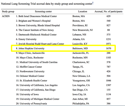
- Primary Result of NLST
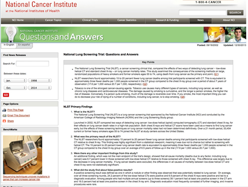
- The NLST researchers found approximately 15 percent to 20 percent fewer lung cancer deaths among trial participants screened with low-dose helical CT relative to chest X-ray. This finding was highly significant from a statistical viewpoint, meaning it was due not to chance but rather to screening with helical CT. The 15 percent to 20 percent lower lung cancer death rate is equivalent to approximately three fewer deaths per 1,000 people screened in the CT group compared to the chest X-ray group over an average of 6.5 years of follow-up in the trial (17.6 per 1,000 versus 20.7 per 1,000). - Secondary Result of NLST
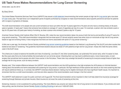
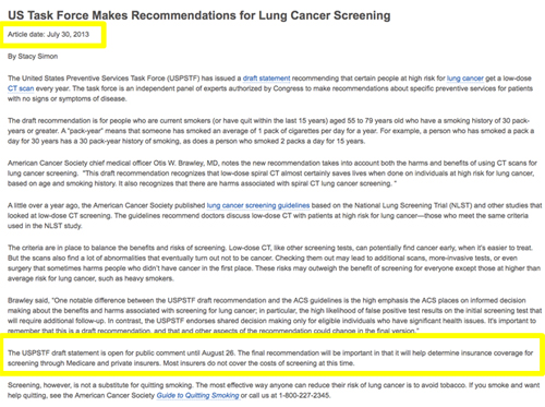
- An additional finding, which was not the main endpoint of the trial's design, showed that all-cause mortality (deaths due to any factor, including lung cancer) was 6.7 percent lower in those screened with low-dose helical CT relative to those screened with chest X-ray. This difference was largely due to the decrease in lung cancer mortality. - “The draft recommendation is for people who are current smokers (or have quit within the last 15 years) aged 55 to 79 years old who have a smoking history of 30 pack-years or greater. A “pack-year” means that someone has smoked an average of 1 pack of cigarettes per day for a year. For example, a person who has smoked a pack a day for 30 years has a 30 pack-year history of smoking, as does a person who smoked 2 packs a day for 15 years.”
- What about false positives?
- A positive screening result was defined as one in which a nodule or other finding was observed that was potentially related to lung cancer. On average, over all three screening rounds, 24.2 percent of the low-dose helical CTs were positive and 6.9 percent of the chest X-rays were positive and led to a diagnostic evaluation. Among people who had multiple annual screens (up to three screens) 39.1 percent had at least one positive screen in the CT arm and 16.0 percent had at least one positive screen in the chest X-ray arm. Diagnostic evaluation most frequently consisted of further imaging, and invasive procedures were rare. - What about false positives?
- Across the three rounds, when a positive screening result was obtained, 96.4 percent of the low-dose helical CT tests and 94.5 percent of the chest X-ray exams were false-positive, meaning that the observed finding was not due to lung cancer. These percentages varied little by round. The vast majority of false-positive results were probably due to the detection of benign lymph nodes or granulomata, which are non-cancerous inflamed tissue masses. The fact that these false-positive results were not cancer was usually confirmed noninvasively by the lack of change in the finding on follow-up CTs. - U.S. Preventive Services Task Force Recommendation Statement 12-31-2013
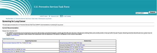
- The USPSTF recommends annual screening for lung cancer with low-dose computed tomography (LDCT) in adults aged 55 to 80 years who have a 30 pack-year smoking history and currently smoke or have quit within the past 15 years. Screening should be discontinued once a person has not smoked for 15 years or develops a health problem that substantially limits life expectancy or the ability or willingness to have curative lung surgery. (B recommendation) - “ Results showed the majority of LDCT screening centers were located in the counties with the highest quartiles of lung cancer incidence and mortality in the Northeast and East North Central states, but several high-risk states had no or few identified screening centers including Oklahoma, Nevada, Mississippi, and Arkansas. As guidelines are implemented and reimbursement for LDCT screening follows, equitable access to LDCT screening centers will become increasingly important, particularly in regions with high rates of lung cancer incidence and smoking prevalence.”
Lung cancer screening using low-dose CT: The current national landscape.
Eberth JM1 et al.
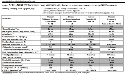
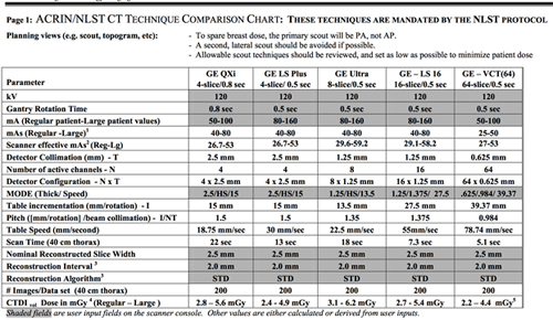
Lung Cancer. 2014 Sep;85(3):379-84 - Low Dose CT Protocols
- NLST Time Capsule
- NLST CT Interpretation
- Negative or minor abnormality: Not suspicious for lung cancer
- Clinically important abnormality: Not suspicious for lung cancer
- Positive: Suspicious for lung cancer - NLST Time Capsule

- CT Screening Scan Protocols 2014
- Our quantitative image analysis indicated that tube current, and thus radiation dose, could be reduced by 40% or 80% from ASIR or MBIR, respectively, compared with conventional FBP, while maintaining similar image noise magnitude and contrast-to-noise ratio.
- Radiation dose reduction for CT lung cancer screening using ASIR and MBIR: a phantom study
- Mathieu KB et al.
- J Applied Clin Med Physics, Vol 15, Number 2, 2014 
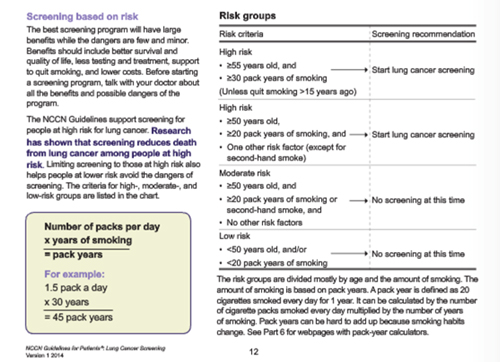
- NCCN Guidelines 2014
- Fleischner society pulmonary nodule recommendations
- Nodule size (mm) less than or equal to 4
- low risk patients - no follow-up needed
- high risk patients - follow-up at 12 months and if no change, no further imaging needed - Fleischner society pulmonary nodule recommendations
- Nodule size > 4-6 mm
- low risk patients - follow-up at 12 months and if no change, no further imaging needed
- high risk patients - initial follow-up CT at 6-12 months and then at 18-24 months if no change - Fleischner society pulmonary nodule recommendations
- Nodule size > 6-8 mm
- low risk patients - initial follow-up CT at 6-12 months and then at 18-24 months if no change
- high risk patients - initial follow-up CT at 3-6 months and then at 9-12 and 24 months if no change - Fleischner society pulmonary nodule recommendations
- Nodule size > 8 mm
- either low or high risk patients
- follow-up CTs at around 3, 9, and 24 months
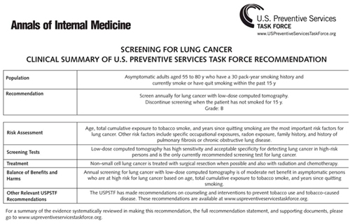
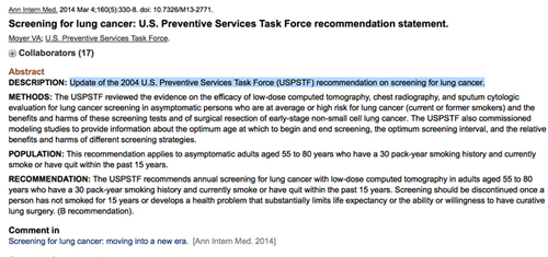
- dynamic contrast enhanced CT, PET, and/or biopsy - “The USPSTF recommends annual screening for lung cancer with low-dose computed tomography in adults aged 55 to 80 years who have a 30 pack-year smoking history and currently smoke or have quit within the past 15 years. Screening should be discontinued once a person has not smoked for 15 years or develops a health problem that substantially limits life expectancy or the ability or willingness to have curative lung surgery. (B recommendation).”
Screening for lung cancer: U.S. Preventive Services Task Force recommendation statement.
Moyer VA et al.
Ann Intern Med. 2014 Mar 4;160(5):330-8 - “This recommendation applies to asymptomatic adults aged 55 to 80 years who have a 30 pack-year smoking history and currently smoke or have quit within the past 15 years.”
Screening for lung cancer: U.S. Preventive Services Task Force recommendation statement.
Moyer VA et al. - Ann Intern Med. 2014 Mar 4;160(5):330-8
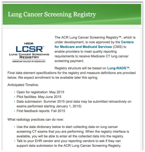
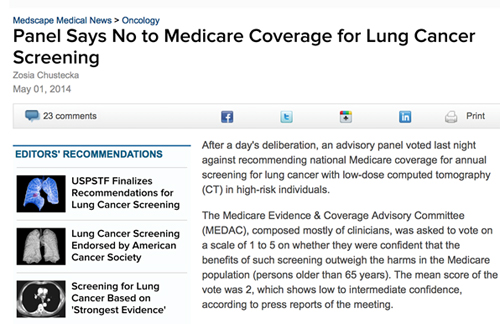
- May 2014 Lung Cancer Screening and Medicare

- “At the T1 and T2 rounds, positive screening results were observed in 27.9% and 16.8% of participants in the low-dose CT group and in 6.2% and 5.0% of participants in the radiography group, respectively. In the low-dose CT group, the sensitivity was 94.4%, the specificity was 72.6%, the positive predictive value was 2.4%, and the negative predictive value was 99.9% at T1; at T2, the positive predictive value increased to 5.2%. In the radiography group, the sensitivity was 59.6%, the specificity was 94.1%, the positive predictive value was 4.4%, and the negative predictive value was 99.8% at T1; both the sensitivity and the positive predictive value increased at T2. Among lung cancers of known stage, 87 (47.5%) were stage IA and 57 (31.1%) were stage III or IV in the low-dose CT group at T1; in the radiography group, 31 (23.5%) were stage IA and 78 (59.1%) were stage III or IV at T1. These differences in stage distribution between groups persisted at T2.”
Results of the two incidence screenings in the National Lung Screening Trial
Aberle DR et al.
N Engl J Med 2013 Sep 5:369(10):920-31 - “At the T1 and T2 rounds, positive screening results were observed in 27.9% and 16.8% of participants in the low-dose CT group and in 6.2% and 5.0% of participants in the radiography group, respectively. In the low-dose CT group, the sensitivity was 94.4%, the specificity was 72.6%, the positive predictive value was 2.4%, and the negative predictive value was 99.9% at T1; at T2, the positive predictive value increased to 5.2%. In the radiography group, the sensitivity was 59.6%, the specificity was 94.1%, the positive predictive value was 4.4%, and the negative predictive value was 99.8% at T1; both the sensitivity and the positive predictive value increased at T2.”
Results of the two incidence screenings in the National Lung Screening Trial
Aberle DR et al.
N Engl J Med 2013 Sep 5:369(10):920-31 - “Among lung cancers of known stage, 87 (47.5%) were stage IA and 57 (31.1%) were stage III or IV in the low-dose CT group at T1; in the radiography group, 31 (23.5%) were stage IA and 78 (59.1%) were stage III or IV at T1. These differences in stage distribution between groups persisted at T2.”
Results of the two incidence screenings in the National Lung Screening Trial
Aberle DR et al.
N Engl J Med 2013 Sep 5:369(10):920-31
- “To conduct a systematic review of the evidence regarding the benefits and harms of lung cancer screening using low-dose computed tomography (LDCT). A multisociety collaborative initiative (involving the American Cancer Society, American College of Chest Physicians, American Society of Clinical Oncology, and National Comprehensive Cancer Network) was undertaken to create the foundation for development of an evidence-based clinical guideline.”
Benefits and harms of CT screening for lung cancer: a systematic review
Bach PB et al.
JAMA 2012 Jun 13;307(22):2418-29 - “Three randomized studies provided evidence on the effect of LDCT screening on lung cancer mortality, of which the National Lung Screening Trial was the most informative, demonstrating that among 53,454 participants enrolled, screening resulted in significantly fewer lung cancer deaths (356 vs 443 deaths; lung cancer?specific mortality, 274 vs 309 events per 100,000 person-years for LDCT and control groups, respectively; relative risk, 0.80; 95% CI, 0.73-0.93; absolute risk reduction, 0.33%; P = .004). The other 2 smaller studies showed no such benefit.”
Benefits and harms of CT screening for lung cancer: a systematic review
Bach PB et al.
JAMA 2012 Jun 13;307(22):2418-29 - “In terms of potential harms of LDCT screening, across all trials and cohorts, approximately 20% of individuals in each round of screening had positive results requiring some degree of follow-up, while approximately 1% had lung cancer. There was marked heterogeneity in this finding and in the frequency of follow-up investigations, biopsies, and percentage of surgical procedures performed in patients with benign lesions. Major complications in those with benign conditions were rare.”
Benefits and harms of CT screening for lung cancer: a systematic review
Bach PB et al.
JAMA 2012 Jun 13;307(22):2418-29 - “Low-dose computed tomography screening may benefit individuals at an increased risk for lung cancer, but uncertainty exists about the potential harms of screening and the generalizability of results.”
Benefits and harms of CT screening for lung cancer: a systematic review
Bach PB et al.
JAMA 2012 Jun 13;307(22):2418-29
“ Low-dose CT screening has been as
sociated with a 20% reduction in lung cancer mortality in a large randomized controlled trial (National Lung Screening Trial [NLST]) of a high-risk population. Mortality data have not yet been reported for 5 other randomized controlled trials, and the sample sizes were too small to detect a meaningful difference in 2 other completed trials. A major risk of CT screening is a high false-positive rate, with associated risks and costs associated with follow-up CT scans and the potential for more invasive diagnostic procedures. Published guidelines for screening indicate a consensus that screening may be indicated for individuals who meet entry criteria for the NLST, but some guidelines expand their recommendations for screening beyond these criteria.”
Computed tomography screening for lung cancer
Boiselle PM
JAMA 2013 Mar 20:309(11):1163-70 - “ A major risk of CT screening is a high false-positive rate, with associated risks and costs associated with follow-up CT scans and the potential for more invasive diagnostic procedures. Published guidelines for screening indicate a consensus that screening may be indicated for individuals who meet entry criteria for the NLST, but some guidelines expand their recommendations for screening beyond these criteria.”
Computed tomography screening for lung cancer
Boiselle PM
JAMA 2013 Mar 20:309(11):1163-70 - “Individuals at high risk of lung cancer who meet the criteria for CT screening in published guidelines should participate in an informed and shared decision-making process by discussing the potential benefits, harms, and uncertainties of screening with their physicians.”
Computed tomography screening for lung cancer
Boiselle PM
JAMA 2013 Mar 20:309(11):1163-70 
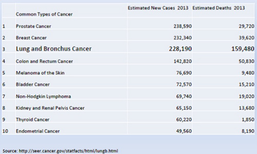
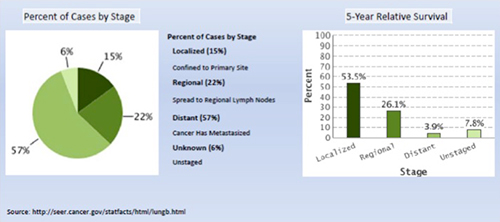
- Lung cancer is the leading cause of cancer-related mortality in the United States and worldwide. In 2012, it is estimated that 160,300 deaths (87,700 in men, 72,600 in women) from lung cancer will occur in the United States. Five-year survival rates for lung cancer are only 15.9%, partly because most patients have advanced-stage lung cancer at initial diagnosis (http://seer.cancer.gov/statfacts/html/lungb.html). lungb.html).
- “The goal of screening is to detect disease at a stage when it is not causing symptoms and when treatment is most successful. Screening should benefit the individual by increasing life expectancy and increasing quality of life. The rate of false-positive results should be low to prevent unnecessary additional testing. The large fraction of the population without the disease should not be harmed (low risk), and the screening test should not be so expensive that it places an onerous burden on the health care system. Thus, the screening test should: 1) improve outcome; 2) be scientifically validated (e.g., have acceptable levels of sensitivity and specificity); and 3) be low risk, reproducible, accessible, and cost effective.”
NCCN Guidelines Version 1.2013 - “The goal of screening is to detect disease at a stage when it is not causing symptoms and when treatment is most successful. Screening should benefit the individual by increasing life expectancy and increasing quality of life. The rate of false-positive results should be low to prevent unnecessary additional testing.”
NCCN Guidelines Version 1.2013 - “The large fraction of the population without the disease should not be harmed (low risk), and the screening test should not be so expensive that it places an onerous burden on the health care system. Thus, the screening test should: 1) improve outcome; 2) be scientifically validated (e.g., have acceptable levels of sensitivity and specificity); and 3) be low risk, reproducible, accessible, and cost effective.”
NCCN Guidelines Version 1.2013 - “It is recommended that institutions performing lung cancer screening use a multidisciplinary approach that may include specialties such as chest radiology, pulmonary medicine, and thoracic surgery. Management of downstream testing and follow-up of small nodules are imperative and may require the establishment of administrative processes to ensure adequate follow-up. Guidelines from ACCP (American College of Chest Physicians) and ASCO (American Society of Clinical Oncology) state that only centers with considerable expertise in lung cancer screening should do LDCT. ”
NCCN Guidelines Version 1.2013 - Who should get screening CT scans?
- Age
- Sex
- Length of time patient smoked
- Amount of smoking (pack years)
- Family history
- Clinical symptoms - NCCN Recommendations
Age 55 to 74 years; 30 or more pack-year history of smoking tobacco; and, if former smoker, have quit within 15 years (category 1). Some high-risk individuals in the NLST also had COPD and other risk factors. This is a category 1 recommendation because these individuals are selected based on the NLST inclusion criteria. An NCCN category 1 recommendation is based on high-level evidence (i.e., randomized controlled trial) and uniform consensus among panel members. Annual screening is recommended for these high-risk individuals for 2 years (category 1) based on the NLST. Annual screening can be considered until the patient is no longer eligible for definitive treatment. However, uncertainty exists about the appropriate duration of screening and the age at which screening is no longer appropriate. - NCCN Recommendations
·Age 50 years or older, 20 or more pack-year history of smoking tobacco, and one additional risk factor (category 2B). This is a category 2B recommendation because these individuals are selected based on lower level evidence (e.g., nonrandomized studies, observational data, and ongoing randomized trials) and because some panel members would not recommend LDCT for these individuals These additional risk factors were previously described and include cancer history, lung disease history, family history of lung cancer, radon exposure, and occupational exposure. Note that the NCCN Lung Cancer Screening Panel does not currently believe that exposure to second-hand smoke is an independent risk factor, because the data are either weak or variable (see “Exposure to Second-Hand Smoke” in this Discussion). - The possible or projected risks of screening for lung cancer using LDCT scans include 1) false-positive results, leading to unnecessary testing, unnecessary invasive procedures (including surgery), increased cost, and decreased quality of life because of mental anguish; 2) false-negative results, which may delay or prevent diagnosis and treatment because of a false sense of good health; 3) futile detection of small aggressive tumors (which have already metastasized, preventing meaningful survival benefit from screening); 4) futile detection of indolent disease (i.e., over diagnosis), which would never have harmed the patient who subsequently undergoes unnecessary therapy; 5) indeterminate results, leading to additional testing; 6) radiation exposure; and 7) physical complications from diagnostic workup. Patients with several comorbid conditions may be at greater risk than those with few or none.
- Potential Risk of Lung Cancer Screening
1) false-positive results, leading to unnecessary testing, unnecessary invasive procedures (including surgery), increased cost, and decreased quality of life because of mental anguish
2) false-negative results, which may delay or prevent diagnosis and treatment because of a false sense of good health;
3) futile detection of small aggressive tumors (which have already metastasized, preventing meaningful survival benefit from screening);
4) futile detection of indolent disease (i.e., overdiagnosis), which would never have harmed the patient who subsequently undergoes unnecessary therapy
5) indeterminate results, leading to additional testing
6) radiation exposure;
7) physical complications from diagnostic workup. Patients with several comorbid conditions may be at greater risk than those with few or none. - “Lung cancer screening with LDCT is a complex and controversial topic, with inherent risks and benefits. Results from the large, prospective, randomized NLST showed that screening with LDCT decreased the relative risk of death from lung cancer by 20% in a select group of high-risk individuals. The NLST results indicate that to prevent one death from lung cancer, 320 high-risk individuals must be screened with LDCT.”
NCCN Guidelines Version 1.2013 Lung Cancer Screening 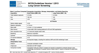
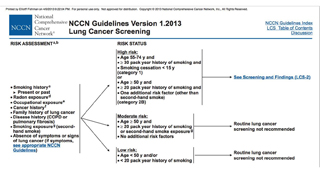
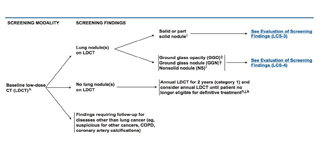
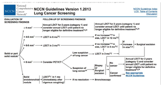
- “After the publication of the NLST (National Lung Screening Trial) results, physicians will be faced with whether to begin ordering low-dose computed tomography (LDCT) of the chest to screen for lung cancer in patients with a history of tobacco use. Despite the encouraging reduction in deaths observed by using LDCT in the NLST study population, recommending adoption of lung cancer screening in general practice is premature. Lessons learned from prostate and breast cancer screening should remind us that the reductions in deaths expected with screening are unfortunately not as readily achievable as initially believed. Furthermore, the potential harms of false-positive findings on chest computed tomography are very real.”
Screening for lung cancer: it works, but does it really work?
Silvestri GA
Ann Intern Med 2011 Oct 18; 155(8):537-9 - “The morbidity and even mortality associated with invasive diagnostic testing and surgical resection due to false- and true-positive findings on computed tomography are likely to increase when the approach taken in the NLST is applied in non-specialty care settings and among the population at highest risk, namely, those with smoking-related comorbid conditions. Although the NLST results are perhaps encouraging, they do not tell us enough that we can be sure that patients who undergo LDCT in an attempt to find early-stage lung cancer will have more benefit than harm.”
Screening for lung cancer: it works, but does it really work?
Silvestri GA
Ann Intern Med 2011 Oct 18; 155(8):537-9 - “ Although the NLST results are perhaps encouraging, they do not tell us enough that we can be sure that patients who undergo LDCT in an attempt to find early-stage lung cancer will have more benefit than harm.”
Screening for lung cancer: it works, but does it really work?
Silvestri GA
Ann Intern Med 2011 Oct 18; 155(8):537-9 - “Screening for lung cancer is not currently recommended, even in persons at high risk for this condition. Most patients with lung cancer present with symptomatic disease that is usually at an incurable, advanced stage. The recently reported NLST (National Lung Screening Trial) showed a 20% decrease in deaths from lung cancer in high-risk persons undergoing screening with low-dose computed tomography of the chest compared with chest radiography. The high-risk group included in the trial comprised asymptomatic persons aged 55 to 74 years, with smoking history of at least 30 pack-years. Screening with low-dose computed tomography detected more cases of early-stage lung cancer and fewer cases of advanced-stage cancer, confirming that screening has shifted the stage of cancer at diagnosis and provides more persons with the opportunity for curative treatment.”
Screening for Lung Cancer: for patients at increased risk for lung cancer,it works
Jett JR, Midthun DE
Ann Intern Med 2011 Oct 18.155(8):540-2 - “ Although computed tomography screening has risks and limitations, the 20% decrease in deaths is the single most dramatic decrease ever reported for deaths from lung cancer, with the possible exception of smoking cessation. Physicians should offer computed tomography screening for lung cancer to patients who fit the high-risk profile defined in the NLST.”
Screening for Lung Cancer: for patients at increased risk for lung cancer,it works
Jett JR, Midthun DE
Ann Intern Med 2011 Oct 18.155(8):540-2 - “Physicians should offer computed tomography screening for lung cancer to patients who fit the high-risk profile defined in the NLST.”
Screening for Lung Cancer: for patients at increased risk for lung cancer,it works
Jett JR, Midthun DE
Ann Intern Med 2011 Oct 18.155(8):540-2 - “On low-dose CT at baseline as compared to CXR, NCNs were detected three times as commonly (23% vs. 7%), malignancies four times as commonly (2.7% vs. 0.7%), Stage I malignancies six times as commonly (2.3% vs. 0.4%). Of the 27 CT-detected cancers, 96% (26/27) were resectable; 85% (23/27) were Stage I, 19 (83%) of the 23 were not seen on CXR. Following the ELCAP recommendations, biopsies were performed on 28 of the 233 subjects with NCNs; 27 had a malignant NCN and one had a benign one. Another three individuals underwent biopsy outside of the ELCAP recommendations, all had benign NCNs. No one had thoracotomy for a benign nodule.”
Early lung cancer action project:overall design and findings from baseline screening
Henschke CI
Cancer 2000 Dec 1;89(11 Suppl) 2474-82 - “On low-dose CT at baseline as compared to CXR, NCNs were detected three times as commonly (23% vs. 7%), malignancies four times as commonly (2.7% vs. 0.7%), Stage I malignancies six times as commonly (2.3% vs. 0.4%). Of the 27 CT-detected cancers, 96% (26/27) were resectable; 85% (23/27) were Stage I, 19 (83%) of the 23 were not seen on CXR.”
Early lung cancer action project:overall design and findings from baseline screening
Henschke CI
Cancer 2000 Dec 1;89(11 Suppl) 2474-82 - “The estimated five-year survival rate of baseline CT-detected malignancies of 60%-80% is a marked improvement over the current rate of 15%. Although false-positive CTs are common, they can be managed with minimal use of invasive diagnostic procedures.”
Early lung cancer action project:overall design and findings from baseline screening
Henschke CI
Cancer 2000 Dec 1;89(11 Suppl) 2474-82 - “After the publication of the NLST (National Lung Screening Trial) results, physicians will be faced with whether to begin ordering low-dose computed tomography (LDCT) of the chest to screen for lung cancer in patients with a history of tobacco use. Despite the encouraging reduction in deaths observed by using LDCT in the NLST study population, recommending adoption of lung cancer screening in general practice is premature. Lessons learned from prostate and breast cancer screening should remind us that the reductions in deaths expected with screening are unfortunately not as readily achievable as initially believed. Furthermore, the potential harms of false-positive findings on chest computed tomography are very real.”
Screening for lung cancer: it works, but does it really work?
Silvestri GA
Ann Intern Med 2011 Oct 18; 155(8):537-9 - “The morbidity and even mortality associated with invasive diagnostic testing and surgical resection due to false- and true-positive findings on computed tomography are likely to increase when the approach taken in the NLST is applied in non-specialty care settings and among the population at highest risk, namely, those with smoking-related comorbid conditions. Although the NLST results are perhaps encouraging, they do not tell us enough that we can be sure that patients who undergo LDCT in an attempt to find early-stage lung cancer will have more benefit than harm.”
Screening for lung cancer: it works, but does it really work?
Silvestri GA
Ann Intern Med 2011 Oct 18; 155(8):537-9 - “ Although the NLST results are perhaps encouraging, they do not tell us enough that we can be sure that patients who undergo LDCT in an attempt to find early-stage lung cancer will have more benefit than harm.”
Screening for lung cancer: it works, but does it really work?
Silvestri GA
Ann Intern Med 2011 Oct 18; 155(8):537-9 - “Screening for lung cancer is not currently recommended, even in persons at high risk for this condition. Most patients with lung cancer present with symptomatic disease that is usually at an incurable, advanced stage. The recently reported NLST (National Lung Screening Trial) showed a 20% decrease in deaths from lung cancer in high-risk persons undergoing screening with low-dose computed tomography of the chest compared with chest radiography. The high-risk group included in the trial comprised asymptomatic persons aged 55 to 74 years, with smoking history of at least 30 pack-years. Screening with low-dose computed tomography detected more cases of early-stage lung cancer and fewer cases of advanced-stage cancer, confirming that screening has shifted the stage of cancer at diagnosis and provides more persons with the opportunity for curative treatment.”
Screening for Lung Cancer: for patients at increased risk for lung cancer,it works
Jett JR, Midthun DE
Ann Intern Med 2011 Oct 18.155(8):540-2 - “ Although computed tomography screening has risks and limitations, the 20% decrease in deaths is the single most dramatic decrease ever reported for deaths from lung cancer, with the possible exception of smoking cessation. Physicians should offer computed tomography screening for lung cancer to patients who fit the high-risk profile defined in the NLST.”
Screening for Lung Cancer: for patients at increased risk for lung cancer,it works
Jett JR, Midthun DE
Ann Intern Med 2011 Oct 18.155(8):540-2 - “Physicians should offer computed tomography screening for lung cancer to patients who fit the high-risk profile defined in the NLST.”
Screening for Lung Cancer: for patients at increased risk for lung cancer,it works
Jett JR, Midthun DE
Ann Intern Med 2011 Oct 18.155(8):540-2 - “There remain unresolved issues with respect to CT screening for lung cancer. These include its feasibility, psychosocial and cost-effectiveness in the UK, harmonisation of CT acquisition techniques, management of suspicious screening findings, the choice of screening frequency and the selection of an appropriate risk group for the intervention. UKLS is aimed at resolving these issues.”
CT screening for lung cancer in the UK: position statement by UKLS investigators following the NLST report
Field JK et al.
Thorax 2011 Aug 66(8):736-7 - NLST (National Lung Screening Trial)
“The aggressive and heterogeneous nature of lung cancer has thwarted efforts to reduce mortality from this cancer through the use of screening. The advent of low-dose helical computed tomography (CT) altered the landscape of lung-cancer screening, with studies indicating that low-dose CT detects many tumors at early stages. The National Lung Screening Trial (NLST) was conducted to determine whether screening with low-dose CT could reduce mortality from lung cancer.” - NLST (National Lung Screening Trial)
“The National Lung Screening Trial (NLST) was conducted to determine whether screening with low-dose CT could reduce mortality from lung cancer. - “ From August 2002 through April 2004, we enrolled 53,454 persons at high risk for lung cancer at 33 U.S. medical centers. Participants were randomly assigned to undergo three annual screenings with either low-dose CT (26,722 participants) or single-view posteroanterior chest radiography (26,732). Data were collected on cases of lung cancer and deaths from lung cancer that occurred through December 31, 2009.”
“ The rate of adherence to screening was more than 90%. The rate of positive screening tests was 24.2% with low-dose CT and 6.9% with radiography over all three rounds. A total of 96.4% of the positive screening results in the low-dose CT group and 94.5% in the radiography group were false positive results. The incidence of lung cancer was 645 cases per 100,000 person-years (1060 cancers) in the low-dose CT group, as compared with 572 cases per 100,000 person-years (941 cancers) in the radiography group (rate ratio, 1.13; 95% confidence interval [CI], 1.03 to 1.23).”
Reduced lung-cancer mortality with low-dose computed tomographic screening
Aberle DR et al.
N Engl J Med 2011 Aug 4;365(5):395-409 - “There were 247 deaths from lung cancer per 100,000 person-years in the low-dose CT group and 309 deaths per 100,000 person-years in the radiography group, representing a relative reduction in mortality from lung cancer with low-dose CT screening of 20.0% (95% CI, 6.8 to 26.7; P=0.004). The rate of death from any cause was reduced in the low-dose CT group, as compared with the radiography group, by 6.7% (95% CI, 1.2 to 13.6; P=0.02).”
Reduced lung-cancer mortality with low-dose computed tomographic screening
Aberle DR et al.
N Engl J Med 2011 Aug 4;365(5):395-409 - “In the NLST, a 20.0% decrease in mortality from lung cancer was observed in the low-dose CT group as compared with the radiography group. The rate of positive results was higher with low-dose CT screening than with radiographic screening by a factor of more than 3, and low-dose CT screening was associated with a high rate of false positive results.”
Reduced lung-cancer mortality with low-dose computed tomographic screening
Aberle DR et al.
N Engl J Med 2011 Aug 4;365(5):395-409 - “Screening with the use of low-dose CT reduces mortality from lung cancer.”
Reduced lung-cancer mortality with low-dose computed tomographic screening
Aberle DR et al.
N Engl J Med 2011 Aug 4;365(5):395-409 - NLST Eligibility for Screening CT/CXR
- Age 55-74 years at time of randomization
- Minimum of 30 pack years, and if former smoker had quit within the 15 prior years
- Prior history of lung cancer, chest CT within 18 months prior to enrollment, had hemoptysis or weight loss over 15 lbs were excluded
- 53,454 enrolled, 26,722 had low dose chest CT, 26733 had radiography at 33 sites nationwide
- Three screenings at 1 yr intervals - CT Screening: Downside
- High false positive rate
- Overdiagnosis (detect lesions which may never have become sympptomatic)
- Potential risk of radiation on f/u screenings
- Risk to patient with workup (i.e. bx) of incidental lesion that is benign - “Although some agencies and organizations are contemplating the establishment of lung-cancer screening recommendations on the basis of the findings of the NLST, the current NLST data alone are, in our opinion, insufficient to fully inform such important decisions.”
Reduced lung-cancer mortality with low-dose computed tomographic screening
Aberle DR et al.
N Engl J Med 2011 Aug 4;365(5):395-409 - “Before public policy recommendations are crafted, the cost-effectiveness of low-dose CT screening must be rigorously analyzed. The reduction in lung-cancer mortality must be weighed against the harms from positive screening results and overdiagnosis, as well as the costs. The cost component of low-dose CT screening includes not only the screening examination itself but also the diagnostic follow-up and treatment.”
Reduced lung-cancer mortality with low-dose computed tomographic screening
Aberle DR et al.
N Engl J Med 2011 Aug 4;365(5):395-409 - October 26,2011
NCCN Guidelines (National Comprehensive Cancer Network)
“Perhaps the most difficult aspect of lung cancer screening is addressing the moral obligation. As part of the Hippocratic oath, physicians promise to first “do no harm.” The dilemma is that if lung cancer screening is beneficial but physicians do not use it, they are denying patients effective care. However, if lung cancer screening is not effective, then patients may be harmed by overdiagnosis, increased testing, invasive testing or procedures, and the anxiety of a potential cancer diagnosis 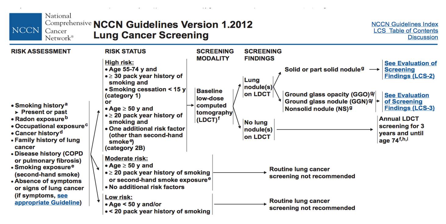
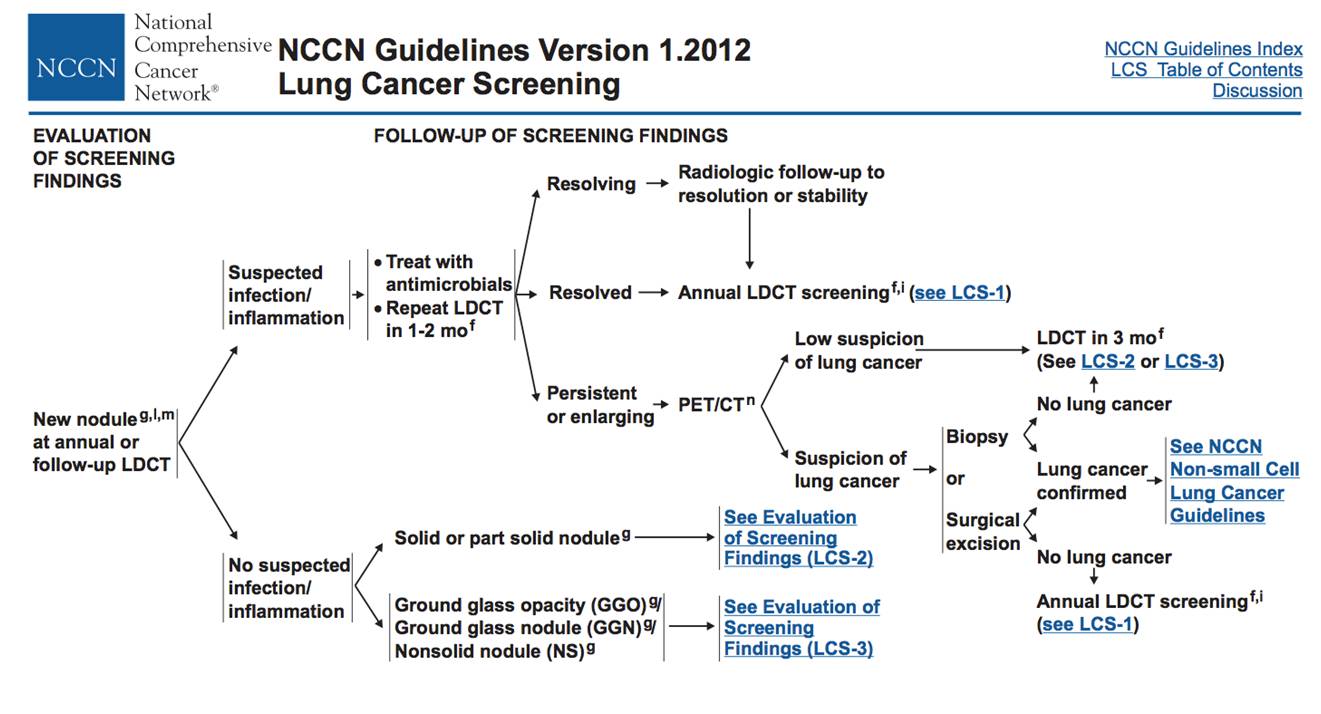
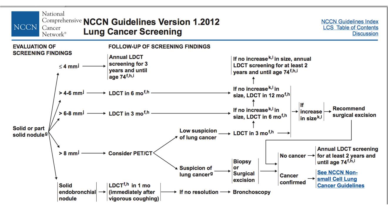
- “Lung cancer screening with CT should be part of a program of care and should not be performed in isolation as a free standing test. Given the high percentage of false-positive results and the downstream management that ensues for many patients, the risks and benefits of lung cancer screening should be discussed with the individual before doing a screening LDCT scan. It is recommended that institutions performing lung cancer screening use a multidisciplinary approach that may include specialties such as radiology, pulmonary medicine, internal medicine, thoracic oncology, and thoracic surgery. Management of downstream testing and follow-up of small nodules are imperative and may require establishment of administrative processes to ensure the adequacy of follow-up.”
- “Lung cancer screening with CT should be part of a program of care and should not be performed in isolation as a free standing test. Given the high percentage of false-positive results and the downstream management that ensues for many patients, the risks and benefits of lung cancer screening should be discussed with the individual before doing a screening LDCT scan.”
- “It is recommended that institutions performing lung cancer screening use a multidisciplinary approach that may include specialties such as radiology, pulmonary medicine, internal medicine, thoracic oncology, and thoracic surgery. Management of downstream testing and follow-up of small nodules are imperative and may require establishment of administrative processes to ensure the adequacy of follow-up.”
- “Screening is recommended (category 1) for high-risk individuals: age 55-74 years; ≥30 pack-year history of smoking tobacco; and if former smoker, have quit within 15 years.7,8 Some high-risk individuals in the NLST also had COPD and other risk factors. This is a category 1 recommendation, because these individuals are selected based on the NLST inclusion criteria.”
- “Annual screening is recommended for these high-risk individuals until they are 74 years old based on the NLST. However, there is uncertainty about the appropriate duration of screening and the age at which screening is no longer appropriate.”
- Screening is also recommended (category 2B) for high-risk individuals: age ≥50 years, ≥20 pack-year history of smoking tobacco, and one additional risk factor. This is a category 2B recommendation from the NCCN panel, because these individuals are selected based on non-randomized studies and observational data. These additional risk factors were previously described and include: cancer history, lung disease history, family history of lung cancer, radon exposure, and occupational exposure.
- Moderate-Risk Individuals
NCCN defines moderate-risk individuals as those: age ≥50 years and ≥20 pack-year history of smoking tobacco or second-hand smoke exposure, but no additional lung cancer risk factors. The NCCN Lung Cancer Screening panel does not recommend lung cancer screening for these moderate-risk individuals. This is a category 2A recommendation, based on non-randomized studies and observational data. - Low-Risk Individuals
NCCN defines low-risk individuals as those: age <50 years and/or smoking history <20 pack-years. The NCCN Lung Cancer Screening panel does not recommend lung cancer screening for these low-risk
individuals. This is a category 2A recommendation, based on non-randomized studies and observational data. - “The NCCN Lung Cancer Screening panel recommends helical LDCT screening for select patients at high risk for lung cancer based on the NLST results, non-randomized studies, and observational data.”
NCCN Guidelines Version 1.2012 Lung Cancer Screening
10-26-2011 - “Lung cancer screening with LDCT is a complex and controversial topic with inherent risks and benefits. Results from the large, prospective, randomized NLST show that lung cancer screening with LDCT can decrease lung cancer specific mortality by 20% and even decrease all-cause mortality by 7%. The NLST results indicate that to prevent one death from lung cancer, 320 high-risk individuals need to be screened with LDCT.”
NCCN Guidelines Version 1.2012 Lung Cancer Screening
10-26-2011 - - 1326 consecutive patients underwent EBCT coronary artery scoring exams
- 25 % former or current smokers
- 2 Board -certified CT radiologists reviewed examinations on a workstation using mediastinal windows, lung windows and bone windows
- Significant extra-cardiac abnormalities were noted
- Horton, Circulation 2002;106:532-534. - - Findings
- 103/1326 patients had extracardiac pathology requiring clinical or imaging follow-up
- 53 patients with noncalcified nodules < 1 cm
- 12 patients with noncalcified nodules > 1 cm
- 24 patients with infiltrates
- 7 patients with indeterminate liver lesions
- 2 patients with sclerotic bone lesions
- 2 patients with breast findings
- 1 patient with polycystic liver disease
- 1 patient with esophageal thickening
- 1 patient with ascites
- Horton, Circulation 2002;106:532-534 - - 503 patients underwent cardiac imaging with 16 or 64 MDCT
- 53% current or former smokers
- Contrast enhanced scans
- Cardiologists assessed the heart
- Radiologists reviewed the other organs
- Reconstructed both large FOV and small FOV
- Onuma JACC 2006;48:402-406. - - Findings
- 346 noncardiac findings were identified in 292 patients (58.1%)
- 114 patients (22.7%) had clinically significant findings
- 3.5% had therapeutic consequences
- 4 cases of malignancy (0.8%)- 2 lung cancers, 1 breast
- 49 patients with noncalcified nodules < 1 cm
- 12 patients with noncalcified nodules > 1 cm
- 16 patients with infiltrates
- 17 patients with pleural effusions
- 7 patients with aortic aneurysms
- Onuma JACC 2006;48:402-406 - - Findings
- 32/ 201 patients in whom coronary disease was ruled out, non cardiac findings by CT were considered sufficient to explain the symptoms
- Onuma JACC 2006;48:402-406. - - 166 patients for contrast enhanced CTA
- Prospective Study
- 16 slice MDCT
- 1mm slices
- Radiologist reviewed soft tissue, lung, and bone windows
- Haller AJR 2006; 187:105-110. - "Extracardiac findings were detected in 41 patients (24.7%). Findings were classified as minor (19.9%) or major (4.8%). Among the major findings, which had an immediate impact on patient management and treatment, were bronchial carcinoma and pulmonary emboli."
Haller
AJR 2006; 187:105-110. - Budoff Cardiovasc Interv 2006; 68:965-73.
- High risk of nodule detection in this population
- Concern with the cost of following these nodules, the radiation dose to the patient, as well as the potential risk of biopsy, etc
- Concern for potential increased cancer risk in patients undergoing follow-up CT scans.
- Concern about unnecessary anxiety for both the patients and physician regarding the follow-up of insignificant findings - "At reduced radiation exposure, low kilovoltage scanning increases the percentage of central and peripheral pulmonary arteries that can be evaluated with CT angiography without a substantial decrease in image quality."
CT Angiography of pulmonary Arteries to Detect Pulmonary Embolism: Improvement of Vascular Enhancement with Low Kilovoltage Settings
Schueller-Weidekamm C et al.
Radiology 2006; 241:899-907.


