|
-- OR -- |
|
- “Coronary artery plaque can consist of calcified and/or noncalcified material. Computed tomography (CT) can detect and measure coronary artery calcium (CAC) levels in calcified plaque, which increases with age and is more common at all ages in men than women. In a study of 19 725 asymptomatic individuals aged 30 to 45 years without known coronary disease from 3US studies, 21% had any detectable CAC (CAC score >0).3 In the Multi-Ethnic Study of Atherosclerosis (MESA) of 6110 asymptomatic US individuals without known cardiovascular disease aged 45 to 84 years (mean age, 62 years), the presence and amount of CAC was greater in older adults. Amon men and women younger than 55 years, more than half of asymptomatic individuals had no detectable CAC. However, among those 80 years or older, more than 80% had some detectable CAC.”
Cardiac CT Calcium Score
Peter Glynn, MD, MSc; Sadiya S. Khan, MD, MSc; Philip Greenland, MD
JAMA. 2025 Mar 5. doi: 10.1001/jama.2025.0610. Online ahead of print. - The standard method to detect and quantify CAC is gated cardiac CT, which uses the patient’s electrocardiogram(ECG) to time CT imaging acquisition to improve image quality. CAC can also be identified on non–ECG-gated CT chest imaging but may not be as precise as an ECG-gated CT. CAC is quantified by the Agatston score, a measure of total coronary atherosclerotic burden. The Agatston score is the sum of the attenuation (in Hounsfield units) and area of all CAC lesions in all coronary arteries. Scores range from 0 (no calcified plaque) to more than 1000(extensive calcified atherosclerosis), with no specific maximum score.
Cardiac CT Calcium Score
Peter Glynn, MD, MSc; Sadiya S. Khan, MD, MSc; Philip Greenland, MD
JAMA. 2025 Mar 5. doi: 10.1001/jama.2025.0610. Online ahead of print. - Incidentally detected CAC on nongated CT chest scans, which is generally reported qualitatively as absent, mild, moderate, or severe, also predicts coronary heart disease (CHD) risk. In patients referred for lung cancer screening due to tobacco use in the National Lung Screening Trial, CAC was present in more than 70% of individuals on nongated thoracic CT scans. In multivariable scores of 1 to100, 101 to1000,andgreater than1000 Agatston units were associated with hazard ratios of 1.27 (95%CI,0.69-2.53), 3.57 (95%CI, 2.14-7.48), and 6.63 (95%CI, 3.57-14.97), respectively.6 analysis of time to CHD death, compared with a CAC score of 0,CAC.
Cardiac CT Calcium Score
Peter Glynn, MD, MSc; Sadiya S. Khan, MD, MSc; Philip Greenland, MD
JAMA. 2025 Mar 5. doi: 10.1001/jama.2025.0610. Online ahead of print. - The most common clinical scenario in which CAC testing should be considered is in an asymptomatic patient undergoing risk assessment for ASCVD. The 2019 ACC/AHA Primary Prevention Guidelines stated that the main purpose of CAC measurement is to facilitate decision-making regarding statin therapy. Therefore, CAC testing is not recommended or individuals already taking a statin, those with known ASCVD, or for individuals with certain ASCVD risk factors (eg, familial hyperlipidemia, diabetes, current smoking) in whom statin therapy is recommended regardless of CAC score.”
Cardiac CT Calcium Score
Peter Glynn, MD, MSc; Sadiya S. Khan, MD, MSc; Philip Greenland, MD
JAMA. 2025 Mar 5. doi: 10.1001/jama.2025.0610. Online ahead of print. - For asymptomatic individuals, CAC testing is not covered by the Centers for Medicare & Medicaid Services and is typically not covered by private insurance. Out-of-pocket costs range from approximately $50 to $400. CAC scoring can also lead to increased costs from additional cardiac testing (stress testing, invasive angiography) and from evaluation of incidental findings (lung nodules, aortic dilation). However, the discovery of a high CAC score in an asymptomatic patient should not automatically lead to additional cardiac testing, since additional testing should be based on the presence of symptoms and any other high risk factors. Recently, many health care systems have made CAC testing available by self referral. Because of the increasing prevalence of self-referral, it will be important to evaluate characteristics of those who self-refer for CAC scoring, and the risk reclassification, downstream testing, and cost-effectiveness in this population.
Cardiac CT Calcium Score
Peter Glynn, MD, MSc; Sadiya S. Khan, MD, MSc; Philip Greenland, MD
JAMA. 2025 Mar 5. doi: 10.1001/jama.2025.0610. Online ahead of print. - “The CAC score improves ASCVD risk assessment, particularly in individuals at borderline or intermediate ASCVD risk who are not already taking statin medication (Table). A CAC score greater than 0 indicates the presence of calcified coronary atherosclerosis and portends higher ASCVD risk compared with those with a CAC score of 0. Statin therapy is recommended for asymptomatic individuals with a CAC score of 100 or greater Agatston units and may be considered for those with a CAC score of 1 to 99 Agatston units, especially in patients younger than 45 years.”
Cardiac CT Calcium Score
Peter Glynn, MD, MSc; Sadiya S. Khan, MD, MSc; Philip Greenland, MD
JAMA. 2025 Mar 5. doi: 10.1001/jama.2025.0610. Online ahead of print. - “As measuring CAC score has become more widely available, this article focuses on 3 situations where CAC testing may be omitted or deferred until a time when CAC testing can provide clinically useful information. Three clinical scenarios to facilitate the clinician-patient risk discussion are as follows: (1) when CAC testing is too early, (2) when CAC testing is too late, and (3) when CAC testing is repeated too often. The timing of CAC testing sits within the decision point of lipid-lowering therapy use. High-risk young adults may face an elevated lifetime risk of cardiovascular disease despite a CAC level of 0, whereas older adults may not see an expected benefit over a short time horizon or may already be taking lipid-lowering therapy, rendering a CAC score less valuable. Integrating a CAC score into the decision to initiate lipid-lowering therapy requires understanding of a patient’s risk factors, including age, as well as the natural history of atherosclerosis and related events.” .
Coronary Artery Calcium Testing-Too Early, Too Late, Too Often.
Zheutlin AR, Chokshi AK, Wilkins JT, Stone NJ
JAMA Cardiol. 2025 Mar 5. doi: 10.1001/jamacardio.2024.5644. Epub ahead of print. PMID: 40042828. - “Repeat imaging must be paired with consideration of how this would change management. The Society of Cardiovascular Computed Tomography expert consensus recommends that repeat imaging may be beneficial among adults with a CAC score of 0 or CAC score greater than 0 and less than 100 in 5 years or 3 to 5 years, respectively. The 2018AHA-ACC-MS guidelines provide a slightly longer range of 5 to 10 years between repeat imaging as a reasonable interval before measuring another CAC score if the initial CAC score was 0 and the patient has no major risk factors (ie, smoking or incident diabetes).” .
Coronary Artery Calcium Testing-Too Early, Too Late, Too Often.
Zheutlin AR, Chokshi AK, Wilkins JT, Stone NJ
JAMA Cardiol. 2025 Mar 5. doi: 10.1001/jamacardio.2024.5644. Epub ahead of print. PMID: 40042828. - “CAC represents overt atherosclerotic disease and conveys more than the probability of disease unlike a calculated risk score. After statin initiation, repeated CAC imaging is not warranted to guide titration of statin therapy. Clinicians should explain to patients why a CAC score in a statin-treated patient does not predict plaque composition as it does for a statin naive patient with a similar score. A CAC score is an important decision tool, and CAC testing should be performed to help guide patient- clinician decisions. However, without considering when it is too early, too late to be useful, or too often (eg, yearly), an opportunity is missed to use this important tool selectively, focusing on those who would most benefit.” .
Coronary Artery Calcium Testing-Too Early, Too Late, Too Often.
Zheutlin AR, Chokshi AK, Wilkins JT, Stone NJ
JAMA Cardiol. 2025 Mar 5. doi: 10.1001/jamacardio.2024.5644. Epub ahead of print.
- ”Coronary fistula (CAF) is a rare coronary artery abnormality in which abnormal blood vessels originating in the coronary arteries bypass the normal capillary and have abnormal communication between the ventricles or large vessels . CAF is mostly congenital and partly acquired, which has been re- ported to occur in approximately 0.1%-0.2% of all patients undergoing invasive coronary angiography . In addition, the prevalence of CAF has been reported to be as high as 0.9% when incidentally detected in coronary computed tomography angiography (CCTA), which has been widely used in recent year . ”
Cinematic rendering of the coronary-pulmonary arterial fistula
Qiang Liu,Jicheng Jiang
R a d i o l o g y Cas e R e p o r t s 1 8 ( 2 0 2 3 ) 3 1 4 0 – 3 1 4 4 - ”Coronary-pulmonary arterial fistula (CPAF) is a type of CAF, which refers to a coronary fistula that terminates at the pulmonary artery. In adults, CPAF is the most common type of CAF, accounting for 76.8%-89.5% of cases. In contrast, coronary-pulmonary fistulas are rare in children, and only 7.5% of patients with coronary-pulmonary fistulas are younger than 18 years of age . Some CPAFs are incidental findings with no clinical significance, but there are some cases with significant hemodynamic effects that require surgical intervention. ”
Cinematic rendering of the coronary-pulmonary arterial fistula
Qiang Liu,Jicheng Jiang
R a d i o l o g y Cas e R e p o r t s 1 8 ( 2 0 2 3 ) 3 1 4 0 – 3 1 4 4 - ”Cinematic rendering, a novel 3D visualization technology for postprocessing of CT images, provides photorealistic images with the potential to improve visualization of anatomic details. Com- pared with traditional volume rendering, cinematic render- ing uses more complex lighting models, enhances surface de- tails, produces realistic shadows, increases the depth of 3D visualization, which can have better visualization effects on the details of the heart structure and coronary vessels [13] . In this case, the global lighting model of cinematic render- ing takes into account the multiple scattering and reflection of the light by the anatomical structures, and the anatomical details and spatial relationships of small blood vessels are highlighted. ”
Cinematic rendering of the coronary-pulmonary arterial fistula
Qiang Liu,Jicheng Jiang
R a d i o l o g y Cas e R e p o r t s 1 8 ( 2 0 2 3 ) 3 1 4 0 – 3 1 4 4
- “In the ED setting, the high accuracy of coronary CTA is driven by its high sensitivity and negative predictive value for detecting or excluding obstructive coronary artery disease, particularly among patients with low-to-intermediate pretest risk for ACS.19–22 In the Perfusion Imaging and CT-Understanding Relative Efficacy (PICTURE) trial,23 coronary CTA demonstrated per-patient sensitivity of 92% and specificity of 87% for significant coronary stenosis using invasive catheter angiography (ICA) as a reference standard, compared with stress myocardial perfusion imaging(per-patient sensitivity of 55% and specificity of 78%).”
2022 use of coronary computed tomographic angiography for patients presenting with acute chest pain to the emergency department: An expert consensus document of the Society of cardiovascular computed tomography (SCCT)
Christopher D. Maroules et al.
Journal of Cardiovascular Computed Tomography 17 (2023) 146–163 - “More recently, the CT-COMPARE trial randomized low-to intermediate risk patients with acute chest pain to coronary CTA (n . 322) or exercise ECG (n . 240). Coronary CTA was associated with 20% reduction in hospital costs, and a 34% reduction in length-of-stay (13.5 hrs vs 20.7 hrs, p < 0.0001). The long-term clinical value of coronary CTA in the ED was evaluated in the 2013 CATCH trial, which randomized 600 patients 1:1 to coronary CTA or functional testing and followed patients for a median duration of 18.7 months. Over this interval, the composite endpoint for MACE (including cardiac death, myocardial infarction, hospitalization for unstable angina, and late symptom-driven revascularization) was less frequent in the coronary CTA arm compared to functional testing (5 vs 14 events, p . 0.04).”
2022 use of coronary computed tomographic angiography for patients presenting with acute chest pain to the emergency department: An expert consensus document of the Society of cardiovascular computed tomography (SCCT)
Christopher D. Maroules et al.
Journal of Cardiovascular Computed Tomography 17 (2023) 146–163 - “Detecting early myocardial injury with high sensitivity troponin (hscTn) assays has become more common throughout the U.S. and internationally. High-sensitivity troponins are preferred over prior generations of conventional cardiac troponin (cTn) tests because they enable faster detection or exclusion of myocardial injury and increase diagnostic accuracy for ACS. However, false positive results and borderline results are a concern as many other cardiac and non-cardiac etiologies can account for elevated hs-cTn measurements beyond epicardial CAD, including myocarditis, arrhythmia, cardiotoxicity, chronic kidney disease, heart failure, sepsis, rhabdomyolysis, chronic obstructive pulmonary disease, and pulmonary embolism.”
2022 use of coronary computed tomographic angiography for patients presenting with acute chest pain to the emergency department: An expert consensus document of the Society of cardiovascular computed tomography (SCCT)
Christopher D. Maroules et al.
Journal of Cardiovascular Computed Tomography 17 (2023) 146–163 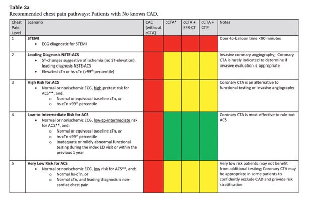
2022 use of coronary computed tomographic angiography for patients presenting with acute chest pain to the emergency department: An expert consensus document of the Society of cardiovascular computed tomography (SCCT)
Christopher D. Maroules et al.
Journal of Cardiovascular Computed Tomography 17 (2023) 146–163- “Coronary CTA is most effective in patients with a low-to-intermediate pretest likelihood of ACS. This includes patients with normal or nonischemic ECG changes, and normal or equivocal baseline cTn, or baseline hs-cTn below the 99th percentile. Using risk stratification tools, low-to-intermediate pretest risk is defined by HEART scores 1–6, and TIMI risk scores 1–4. This category also includes patients with inadequate or mildly abnormal functional testing during the index ED visit or within the previous 1 year.”
2022 use of coronary computed tomographic angiography for patients presenting with acute chest pain to the emergency department: An expert consensus document of the Society of cardiovascular computed tomography (SCCT)
Christopher D. Maroules et al.
Journal of Cardiovascular Computed Tomography 17 (2023) 146–163
- OBJECTIVE. Previously published reports have shown that coronary CT angiography (CCTA) is a more efficient method of diagnosis than myocardial perfusion imaging (MPI) and stress echocardiography for patients presenting to emergency departments (EDs) with acute chest pain. In light of this evidence, the objective of this study was to examine recent trends in the use of these techniques in EDs.
CONCLUSION. Use of CCTA in EDs has increased rapidly, but far more MPI examinations are still being performed. This finding suggests that recently acquired evidence is not yet being fully acted upon.
Coronary CT Angiography: Use in Patients With Chest Pain Presenting to Emergency Departments Levin DC et al. AJR 2018; 210:816–820 - “ Chest pain is the second most com- mon cause of patient visits to emergency departments (EDs) in the United States. There are 8 million such visits annually, and they result in approximately $12 billion in costs . On arrival in the ED, patients with chest pain typically undergo ECG and cardiac enzyme determination. If these tests do not yield a diagnosis of acute coronary syndrome, the patient generally undergoes further observation and repeat testing. The testing often then includes imaging.”
Coronary CT Angiography: Use in Patients With Chest Pain Presenting to Emergency Departments Levin DC et al. AJR 2018; 210:816–820 - “The American College of Radiology Ap- propriateness Criteria include a listing for chest pain suggestive of acute coronary syndrome. These criteria, which are well known to radiologists, rate various imaging tests on a scale of 1–9 with 7–9 considered appropriate, 4–6 possibly appropriate, and 1–3 usually not appropriate. In this particular clinical circumstance, MPI is rated 8, stress echocardiography 7, and CCTA 6. The reasons for the low rating of CCTA are not clear.”
Coronary CT Angiography: Use in Patients With Chest Pain Presenting to Emergency Departments Levin DC et al. AJR 2018; 210:816–820 - “In view of what the aforementioned literature has shown, we believe the appropriateness criteria for chest pain suggestive of acute coronary syndrome should be revised to rate CCTA higher than both MPI and stress echocardiography. Radiologists, who interpret most CCTA examinations performed in EDs, should educate their emergency medicine colleagues about the greater utility of CCTA in the evaluation of patients with chest pain.”
Coronary CT Angiography: Use in Patients With Chest Pain Presenting to Emergency Departments Levin DC et al. AJR 2018; 210:816–820 - “An apparent limitation of CR is illustrated in the 2nd case. While the shadowing effects that arise from the global lighting model that is used contribute to the photorealistic quality of the images, shadowing can also potentially obscure important pathology. Therefore, as with any 3D rendering technique, it is important for the user to examine CR visualizations from multiple different views and to interactively alter the windowing settings to increase and decrease the transparency of overlapping anatomic structures, and to use CR in conjunction with axial display, 2D MPRs, MIP and VR. Other limitations of CR may come to light as the method becomes more widely clinically available.”
Cinematic rendering of cardiac CT volumetric data: Principles and initial observations Steven P. Rowe, Pamela T. Johnson, Elliot K. Fishman Journal of Cardiovascular Computed Tomography 12 (2018) 56–59 - “CR is a promising method to enhance display volumetric CT data and should prove useful in diagnosis, treatment planning, surgical navigation, trainee education, and patient engagement. However, further study is needed to establish the advantaged and disadvantages of CR in comparison to other 3D methods.”
Cinematic rendering of cardiac CT volumetric data: Principles and initial observations Steven P. Rowe, Pamela T. Johnson, Elliot K. Fishman Journal of Cardiovascular Computed Tomography 12 (2018) 56–59
“The intent of CAD-RADS – Coronary Artery Disease Reporting and Data System is to create a standardized method to communicate findings of coronary CT angiography (coronary CTA) in order to facilitate decision-making regarding further patient management.”
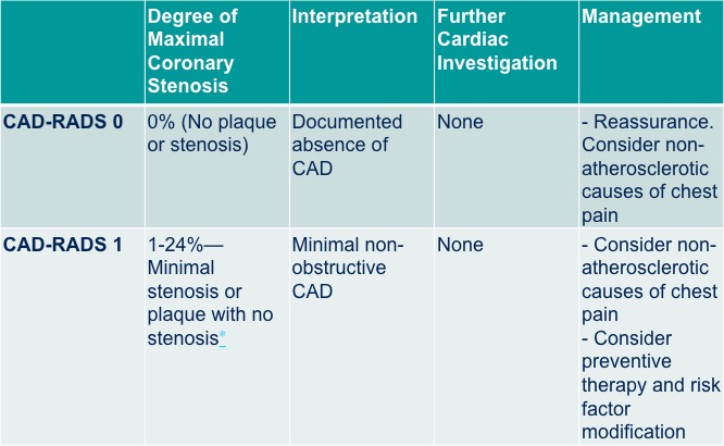
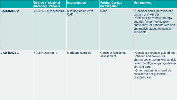
CAD-RADS™: Coronary Artery Disease - Reporting and Data System: An Expert Consensus Document of the Society of Cardiovascular Computed Tomography (SCCT), the American College of Radiology (ACR) and the North American Society for Cardiovascular Imaging (NASCI). Endorsed by the American College of Cardiology. Cury RC et al. J Am Coll Radiol. 2016 Dec;13(12 Pt A):1458-1466.“The suggested CAD-RADS classification is applied on a per-patient basis and represents the highest-grade coronary artery lesion documented by coronary CTA. It ranges from CAD-RADS 0 (Zero) for the complete absence of stenosis and plaque to CAD-RADS 5 for the presence of at least one totally occluded coronary artery and should always be interpreted in conjunction with the impression found in the report. Specific recommendations are provided for further management of patients with stable or acute chest pain based on the CAD-RADS classification.”
CAD-RADS™: Coronary Artery Disease - Reporting and Data System: An Expert Consensus Document of the Society of Cardiovascular Computed Tomography (SCCT), the American College of Radiology (ACR) and the North American Society for Cardiovascular Imaging (NASCI). Endorsed by the American College of Cardiology. Cury RC et al. J Am Coll Radiol. 2016 Dec;13(12 Pt A):1458-1466.“The main goal of CAD-RADS is to standardize reporting of coronary CTA results and to facilitate communication of test results to referring physicians along with suggestions for subsequent patient management. In addition, CAD-RADS will provide a framework of standardization that may benefit education, research, peer-review and quality assurance with the potential to ultimately result in improved quality of care.”
CAD-RADS™: Coronary Artery Disease - Reporting and Data System: An Expert Consensus Document of the Society of Cardiovascular Computed Tomography (SCCT), the American College of Radiology (ACR) and the North American Society for Cardiovascular Imaging (NASCI). Endorsed by the American College of Cardiology. Cury RC et al. J Am Coll Radiol. 2016 Dec;13(12 Pt A):1458-1466.Reporting Standards
BI-RADS (Breast Imaging with standardized reporting of screening mammograms )
LI-RADS™ (Liver Imaging Reporting and Data System) for standardization reporting in patients with chronic liver disease.
Lung-RADS™ (Lung CT Screening Reporting and Data System) for standardization reporting of high-risk smokers undergoing CT lung screening.
PI-RADS™ (Prostate Imaging Reporting and Data System) for multi-parametric MR imaging in the context of prostate cancer.“The goal of CAD-RADS, through standardization of report terminology for coronary CTA, is to improve communication between interpreting and referring physicians, facilitate research, and offer mechanisms to contribute to peer review and quality assurance, ultimately resulting in improvements to quality of care. Importantly, CAD-RADS does not substitute the impression section provided by the reading physician and should always be interpreted in conjunction with the more individual and patient-specific information found in the report.”
CAD-RADS™: Coronary Artery Disease - Reporting and Data System: An Expert Consensus Document of the Society of Cardiovascular Computed Tomography (SCCT), the American College of Radiology (ACR) and the North American Society for Cardiovascular Imaging (NASCI). Endorsed by the American College of Cardiology. Cury RC et J Am Coll Radiol. 2016 Dec;13(12 Pt A):1458-1466.“The main clinical benefit of coronary CTA is derived from its high sensitivity and negative predictive value. The positive predictive value of coronary CTA is lower, and especially intermediate lesions may be overestimated regarding their relevance. Many patients with previously known CAD will include lesions that fall into this category, so that coronary CTA will need to be complemented by further tests.”
CAD-RADS™: Coronary Artery Disease - Reporting and Data System: An Expert Consensus Document of the Society of Cardiovascular Computed Tomography (SCCT), the American College of Radiology (ACR) and the North American Society for Cardiovascular Imaging (NASCI). Endorsed by the American College of Cardiology. Cury RC et J Am Coll Radiol. 2016 Dec;13(12 Pt A):1458-1466.“The use of coronary CTA to assess patients with stable chest pain in the outpatient setting or acute chest pain presenting to the Emergency Department has been validated in various clinical trials. Major guidelines are incorporating the use of coronary CT angiography as appropriate for assessing low to intermediate risk patients presenting with chest pain. Decreasing the variation in reporting is one aspect that will contribute to wider dissemination in clinical practice, minimize error and to ultimately improve patient outcome. The main goal of the CAD-RADS classification system is to propose a reporting structure that provides consistent categories for final assessment, along with suggestions for further management.”
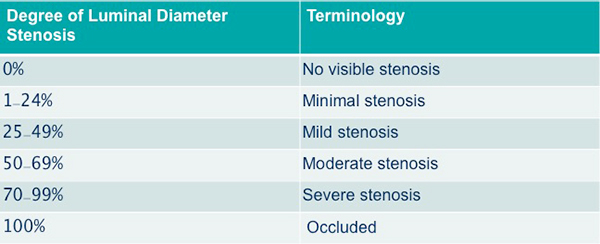
CAD-RADS™: Coronary Artery Disease - Reporting and Data System: An Expert Consensus Document of the Society of Cardiovascular Computed Tomography (SCCT), the American College of Radiology (ACR) and the North American Society for Cardiovascular Imaging (NASCI). Endorsed by the American College of Cardiology. Cury RC et J Am Coll Radiol. 2016 Dec;13(12 Pt A):1458-1466.SCCT Grading for Coronary Artery Stenosis
CAD-RADS Reporting and Data System for patients presenting with stable chest pain
CAD-RADS Reporting and Data System for patients presenting with stable chest pain
CAD-RADS Reporting and Data System for patients presenting with stable chest pain
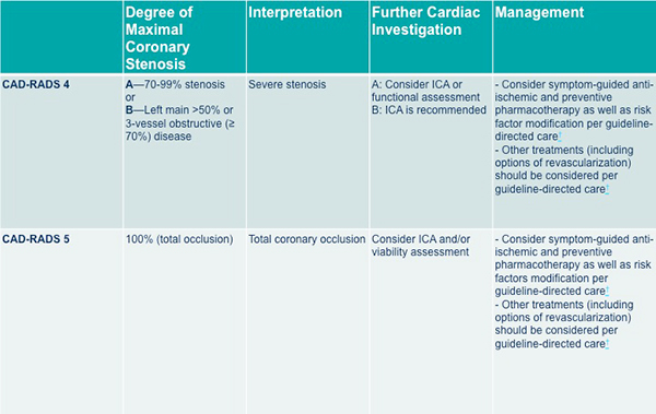
CAD-RADS Reporting and Data System for patients presenting with stable chest pain
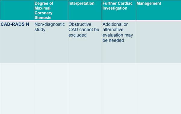
Acute chest pain, negative first troponin, negative or non-diagnostic electrocardiogram and low to intermediate risk (TIMI risk score < 4)
Acute chest pain, negative first troponin, negative or non-diagnostic electrocardiogram and low to intermediate risk (TIMI risk score < 4)
Acute chest pain, negative first troponin, negative or non-diagnostic electrocardiogram and low to intermediate risk (TIMI risk score < 4)
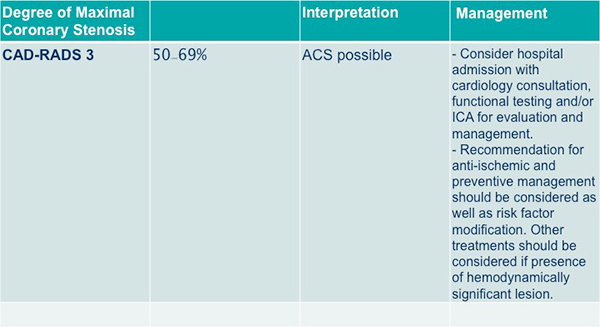
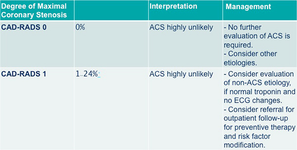
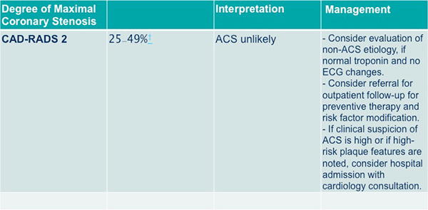
Acute chest pain, negative first troponin, negative or non-diagnostic electrocardiogram and low to intermediate risk (TIMI risk score < 4)
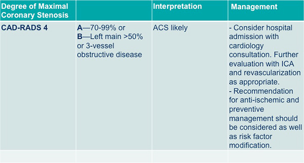
Acute chest pain, negative first troponin, negative or non-diagnostic electrocardiogram and low to intermediate risk (TIMI risk score < 4)
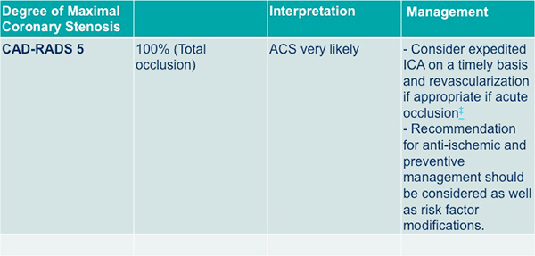
Acute chest pain, negative first troponin, negative or non-diagnostic electrocardiogram and low to intermediate risk (TIMI risk score < 4)

- OBJECTIVE: To evaluate the effectiveness of the iPad (Apple Inc., Cupertino, CA) for two-dimensional (2D) reading of CT angiography (CTA) studies performed for suspected acute non-variceal gastrointestinal bleeding.
CONCLUSION: Compared with a conventional PACS workstation, iPad-based preliminary 2D reading of CTA studies has comparable diagnostic accuracy for detection of acute gastrointestinal bleeding and can be significantly faster.
ADVANCES IN KNOWLEDGE: The iPad could be used by on-call interventional radiologists for immediate decision on percutaneous embolization in patients with suspected acute gastrointestinal bleeding.
iPad-based primary 2D reading of CT angiography examinations of patients with suspected acute gastrointestinal bleeding: preliminary experience.
Faggioni L et al.
Br J Radiol. 2015 Mar;88(1047) - " For both readers, there was no significant difference in agreement with the reference standard for per-vessel stenosis scores using either the 3D workstation or the iPad. In a multivariable logistic regression analysis including reader, workstation, and vessel as co-variates, there was no significant association between workstation type or reader and agreement with the reference standard (p > 0.05). Both readers identified 100 % of coronary anomalies using each technique."
Remote reading of coronary CTA exams using a tablet computer: utility for stenosis assessment and identification of coronary anomalies
Stefan L. Zimmerman, Cheng T. Lin, Linda C. Chu, John Eng, Elliot K. Fishman
Emerg Radiol DOI 10.1007/s10140-016-1399-9 - "However, in many institutions, coronary CTA access is limited to daytime and weekday hours, at least in part due to limits on availability of subspecialty trained physicians and the need for post-processing workstations. Remote reading of coronary CTA studies could expand access by providing a platform for after hour emergency department coverage on an as needed basis without requiring imagers to be in-house."
Remote reading of coronary CTA exams using a tablet computer: utility for stenosis assessment and identification of coronary anomalies
Stefan L. Zimmerman, Cheng T. Lin, Linda C. Chu, John Eng, Elliot K. Fishman
Emerg Radiol DOI 10.1007/s10140-016-1399-9 - "Coronary CTA with using a tablet computer is feasible with results that are no different from reading of cardiac exams on standard clinical workstations. On multivariate analysis, we found no significant relationship between the type of reading modality and accuracy of interpretation. Remote reading with a tablet computer could be used to expand availability of coronary CTA in the ED."
Remote reading of coronary CTA exams using a tablet computer: utility for stenosis assessment and identification of coronary anomalies
Stefan L. Zimmerman, Cheng T. Lin, Linda C. Chu, John Eng, Elliot K. Fishman
Emerg Radiol DOI 10.1007/s10140-016-1399-9 -
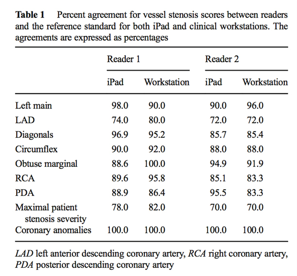
Remote reading of coronary CTA exams using a tablet computer: utility for stenosis assessment and identification of coronary anomalies
Stefan L. Zimmerman, Cheng T. Lin, Linda C. Chu, John Eng, Elliot K. Fishman
Emerg Radiol DOI 10.1007/s10140-016-1399-9
- “With current economic constraints, cost-effective strategies are needed to better risk stratify the patients. CCTA may represent a cost-effective, noninvasive tool for assessment of CAD and may be used as a gatekeeper for costly invasive procedures such as ICA in appropriate circumstances. Hence, further research especially randomized controlled trials are needed to validate the role of CCTA as a cost-effective means of CAD assessment and as a gatekeeper to ICA.” Coronary computed tomography as a cost–effective test strategy for coronary artery disease assessment – A systematic review Zeb I et al. Atherosclerosis Vol 234, Issue 2, June 2014, Pages 426–435
- “There are certain limitations to the use of CCTA as a gatekeeper to ICA. CCTA lacks the ability to provide the functional consequence of the anatomical lesions. There are concerns that the use of CCTA may result in increased radiation exposure for the patient. The current advances in the technology, improvements in image acquisition protocols and post processing of the images including iterative reconstructive has led to decrease in the radiation exposure associated with the use of CCTA. There are certain limitations that are inherent to the use of CCTA that limit the diagnostic accuracy of the test such as coronary artery motion, high CAC and image noise. Local expertise in the evaluation of CCTA is an important factor that significantly contributes to assessing diagnostic accuracy.” Coronary computed tomography as a cost–effective test strategy for coronary artery disease assessment – A systematic review Zeb I et al. Atherosclerosis Vol 234, Issue 2, June 2014, Pages 426–435
- “Recent studies now support the use of statin therapy in patients with CAC>0, and individualization of aspirin considering patient ASCVD risk factors, CAC severity, and factors that predispose to bleeding.” Coronary computed tomography as a cost–effective test strategy for coronary artery disease assessment – A systematic review Zeb I et al. Atherosclerosis Vol 234, Issue 2, June 2014, Pages 426–435
- “It is therefore of utmost importance that interventional cardiologists become familiar with image interpretation and up-to-date regarding several CTA features, taking advantage of this information in planning the procedure, ultimately leading to improvement in patient outcomes. On the other hand, the increasing use of CCTA as a gatekeeper for invasive coronary angiography is expected to lead to an increase in the ratio of interventional to diagnostic procedures and significant changes in the daily cath-lab routine. In a foreseeable future, cath-labs will probably offer an invasive procedure only to patients expected to undergo an intervention, perhaps becoming in this change true interventional-labs.”
Computed tomography angiography for the interventional cardiologist.
de Araújo Gonçalves P et al
Eur Heart J Cardiovasc Imaging. 2014 Aug;15(8):842-54. - “On the other hand, the increasing use of CCTA as a gatekeeper for invasive coronary angiography is expected to lead to an increase in the ratio of interventional to diagnostic procedures and significant changes in the daily cath-lab routine. In a foreseeable future, cath-labs will probably offer an invasive procedure only to patients expected to undergo an intervention, perhaps becoming in this change true interventional-labs.”
Computed tomography angiography for the interventional cardiologist.
de Araújo Gonçalves P et al
Eur Heart J Cardiovasc Imaging. 2014 Aug;15(8):842-54. - “In recent years, coronary CT angiography (CCTA) has become a widely adopted technique, not only due to its high diagnostic accuracy, but also to the fact that CCTA provides a comprehensive evaluation of the total (obstructive and non-obstructive) coronary atherosclerotic burden. More recently, this technique has become mature, with a large body of evidence addressing its prognostic validation. In addition, CT angiography has moved from the field of 'imagers' and clinicians and entered the interventional cardiology arena, aiding in the planning of both coronary and structural heart interventions, being transcatheter aortic valve implantation one of its most successful examples.”
Computed tomography angiography for the interventional cardiologist.
de Araújo Gonçalves P et al
Eur Heart J Cardiovasc Imaging. 2014 Aug;15(8):842-54. - “In recent years, coronary CT angiography (CCTA) has become a widely adopted technique, not only due to its high diagnostic accuracy, but also to the fact that CCTA provides a comprehensive evaluation of the total (obstructive and non-obstructive) coronary atherosclerotic burden. More recently, this technique has become mature, with a large body of evidence addressing its prognostic validation.”
Computed tomography angiography for the interventional cardiologist.
de Araújo Gonçalves P et al
Eur Heart J Cardiovasc Imaging. 2014 Aug;15(8):842-54.
- “ Three large randomized trials (CT-STAT, ACRIN-PA, and ROMICAT II) have compared a coronary CTA strategy with current standard of care evaluations in >3000 patients. These trials consistently show the safety of a negative coronary CT angiogram to identify patients for discharge from the emergency department with low rates of adverse cardiovascular events, at significantly lower cost, and greater efficiency in terms of time to discharge.”
Coronary CT angiography versus standard of care for assessment of chest pain in the emergency department
Cury RC, Budoff M, Taylor AJ
J Cardiovasc Comput Tomogr 7(2013):79-82 - “ Together, these trials provide definitive evidence for the use of coronary CTA in the emergency department in patients with a low to intermediate pretest probability of coronary artery disease. Clinical practice guidelines that recommend the use of coronary CTA in the emergency department are warranted.”
Coronary CT angiography versus standard of care for assessment of chest pain in the emergency department
Cury RC, Budoff M, Taylor AJ
J Cardiovasc Comput Tomogr 7(2013):79-82
- “ Premature CHD is a common cause of death among firefighters with an average age of death of 44 years. Use of cardiac CT in firefighters may provide a more accurate method of risk assessment in this population.”
Coronary calcium scoring independently detects coronary artery disease in asymptomatic firefighters: A prospective study
Santora LJ et al.
J Cardiovasc Comput Tomogr 7 (2013) 46-50 - “ Firefighters have a high burden of calcified coronary atherosclerosis, greater than anticipated on the basis of age and coronary risk factors.”
Coronary calcium scoring independently detects coronary artery disease in asymptomatic firefighters: A prospective study
Santora LJ et al.
J Cardiovasc Comput Tomogr 7 (2013) 46-50
- Advantages and Disadvantages of Cardiac CT
Advantages
- High temporal and spatial resolution
- Three dimensional dataset
- Rapid acquisition
- Wide availability
Complications of Aortic Valve Surgery: Manifestations at CT and MR Imaging
Pham N et al.
RadioGraphics 2012; 32:1873-1892 - Advantages and Disadvantages of Cardiac CT
Disadvantages
- Severe artifacts with certain prosthetic valves (Bjork Shiley)
- Radiation exposure
- Need for iodinated contrast material
- Need for heart rate control
- Irregular heart rhythm may lead to motion artifact (atrial fibrillation and ventricular extrasystoles)
Complications of Aortic Valve Surgery: Manifestations at CT and MR Imaging
Pham N et al.
RadioGraphics 2012; 32:1873-1892
- “ In patients with high likelihood of CAD, the performance of coronary CT angiography in the differentiation of patients without and patients with a need for revascularization and the selection of revascularization strategy was similar to that of cardiac catherization; accordingly, coronary CT angiography has the potential to limit the number of patients without obstructive CAD who undergo cardiac catheterization and to inform decision making regarding revascularization.”
Coronary CT Angiography versus Conventional Cardiac Angiography for Therapeutic Decision Making in Patients with High Likelihood of Coronary Artery Disease
Moscariello A et al
Radiology 2012; 265:385-392 - “ In patients with high likelihood of CAD, the performance of coronary CT angiography in the differentiation of patients without and patients with a need for revascularization and the selection of revascularization strategy was similar to that of cardiac catheterization.”
Coronary CT Angiography versus Conventional Cardiac Angiography for Therapeutic Decision Making in Patients with High Likelihood of Coronary Artery Disease
Moscariello A et al
Radiology 2012; 265:385-392 - “ Coronary CT angiography has the potential to limit the number of patients without obstructive coronary artery disease who undergo conventional cardiac catheterization and to inform decision making with regard to revascularization.”
Coronary CT Angiography versus Conventional Cardiac Angiography for Therapeutic Decision Making in Patients with High Likelihood of Coronary Artery Disease
Moscariello A et al
Radiology 2012; 265:385-392 - “Our findings also confirm the relatively low specificity of clinical decision making based on an abnormal SPECT myocardial perfusion imaging studies, as 59% of patients referred for cardiac catheterization on the basis of positive SPECT studies did not have obstructive CAD.”
Coronary CT Angiography versus Conventional Cardiac Angiography for Therapeutic Decision Making in Patients with High Likelihood of Coronary Artery Disease
Moscariello A et al
Radiology 2012; 265:385-392 - “The performance parameters of coronary CT angiography in this study, with a per-patient sensitivity and specificity of 100% and 93.6%, respectively, are consistent with the results of previous investigations that have demonstrated the strong performance of coronary CT angiography as a noninvasive alternative to conventional cardiac catheterization for the detection and exclusion of obstructive CAD.”
Coronary CT Angiography versus Conventional Cardiac Angiography for Therapeutic Decision Making in Patients with High Likelihood of Coronary Artery Disease
Moscariello A et al
Radiology 2012; 265:385-392
- “ In the setting of undifferentiated chest pain, CT Angiography (CTA) with its high sensitivity and specificity can be considered the modality of choice to diagnose suspected PE or aortic pathology such as aortic dissection or aneurysm.”
ACR Appropriateness Criteria Acute Chest Pain-Low Probability of Coronary Artery Disease
Hoffman U et al.
J Am Coll Radiol 2012;9:745-750 - “ Most important, in this low-risk population, cardiac CTA has nearly perfect negative predictive value to rule out significant CAD. Multidetector CT is also the primary method for diagnosing coronary anomalies, a rare cause of acute chest pain.”
ACR Appropriateness Criteria Acute Chest Pain-Low Probability of Coronary Artery Disease
Hoffman U et al.
J Am Coll Radiol 2012;9:745-750 - “ With advanced CT technology, it is possible to perform a single phase triple rule out examination allowing comprehensive assessment of CAD, aortic dissection, and PE. However, its efficiency or effectiveness has not been demonstrated.”
ACR Appropriateness Criteria Acute Chest Pain-Low Probability of Coronary Artery Disease
Hoffman U et al.
J Am Coll Radiol 2012;9:745-750
- “ Compared to patients with normal sinus rhythm (NSR), patients with atrial fibrillation/flutter had the highest radiation exposure, followed by those with premature atrial contraction /premature ventricular contraction. Even after adjustment for factors associated with radiation exposure a significant difference in radiation dose persisted.”
The effect of heart rhythm on patient radiation dose with dual source cardiac computed tomography
Techasith T et al.
J Cardiovasc Comput Tomogr (2011) 5;255-263 "Performing coronary CTA before cardiac catheterization is a cost effective strategy in the care of patients without symptoms who have positive stress test results when the probability that the patient has significant coronary disease is less than 50%."
Cost Effectiveness of Coronary CT Angiography in Evaluation of Patients Without Symptoms Who Have Positive Stress Test Results
Halpern EJ et al.
AJR 2010; 194:1257-1262"Significant Left main coronary artery disease was found in 2.4% of the 1,000 patients referred to 64 MSCT examinations. Age and male gender were the independent predictors for LMCAD."
Prevalence of left main coronary artery disease among patients referred to multislice computed tomography coronary examinations
Gemici G et al.
Int J Cardiovasc Imaging (2009) 25:433-438- Triage with MDCT
- CCS under 10 stop, over 10 go to CCTA
- CCTA without 50% or greater stenosis stop
- CCTA greater than 50% stenosis goes to catheter angiography "Those with CCS>10 are then evaluated by noninvasive CT angiography (CTA) which has a high sensitivity and specificity for detecting coronary artery stenosis."
Cost-effectiveness of MDCT compared with myocardial perfusion imaging as gatekeeper to invasive coronary angiography in asymptomatic firefighters with positive treadmill tests
Budoff MJ et al
J Cardiovasc Comput Tomogr (2009) 3, 323-330"In this firefighter population, the cost of ICA-confirmed diagnosis of CAD is substantially lower with MDCT as gatekeeper than with MPI (myocardial perfusion imaging)."
Cost-effectiveness of MDCT compared with myocardial perfusion imaging as gatekeeper to invasive coronary angiography in asymptomatic firefighters with positive treadmill tests
Budoff MJ et al
J Cardiovasc Comput Tomogr (2009) 3, 323-330"This study demonstrates that coronary CT angiography is a cost effective method for evaluation of individuals with stable chest pain syndrome and intermediate CAD prevalence in the long term period."
Cost-effectiveness of Coronary CT Angiography versus Myocardial Perfusion SPECT for Evaluation of patients with Chest Pain and No Known Coronary Artery Disease
Min JK et al.
Radiology 2010;254:801-808"With a $20,000 threshold level for cost per correct diagnosis and $50,000 per quality adjusted life year (QALY), a coronary CT angiography-only approach is the most cost effective diagnostic strategy for evaluation of patients who have stable chest pain without known CAD with intermediate CAD prevalence."
Cost-effectiveness of Coronary CT Angiography versus Myocardial Perfusion SPECT for Evaluation of patients with Chest Pain and No Known Coronary Artery Disease
Min JK et al.
Radiology 2010;254:801-808Will Cardiac CTA help with patient selection?
N Engl J Med. 2010 Mar 11;362(10):943-5
New Low Diagnostic Yield of Elective Coronary Angiography
Manesh R. Patel, M.D., Eric D. Peterson, M.D., M.P.H., David Dai, M.S., J. Matthew Brennan, M.D., Rita F. Redberg, M.D., H. Vernon Anderson, M.D., Ralph G. Brindis, M.D., and Pamela S. Douglas, M.D.
ABSTRACT Background Guidelines for triaging patients for cardiac catheterization recommend a risk assessment and noninvasive testing. We determined patterns of noninvasive testing and the diagnostic yield of catheterization among patients with suspected coronary artery disease in a contemporary national sample. Methods From January 2004 through April 2008, at 663 hospitals in the American College of Cardiology National Cardiovascular Data Registry, we identified patients without known coronary artery disease who were undergoing elective catheterization. The patients' demographic characteristics, risk factors, and symptoms and the results of noninvasive testing were correlated with the presence of obstructive coronary artery disease, which was defined as stenosis of 50% or more of the diameter of the left main coronary artery or stenosis of 70% or more of the diameter of a major epicardial vessel. Results A total of 398,978 patients were included in the study. The median age was 61 years; 52.7% of the patients were men, 26.0% had diabetes, and 69.6% had hypertension. Noninvasive testing was performed in 83.9% of the patients. At catheterization, 149,739 patients (37.6%) had obstructive coronary artery disease. No coronary artery disease (defined as <20% stenosis in all vessels) was reported in 39.2% of the patients. Independent predictors of obstructive coronary artery disease included male sex (odds ratio, 2.70; 95% confidence interval [CI], 2.64 to 2.76), older age (odds ratio per 5-year increment, 1.29; 95% CI, 1.28 to 1.30), presence of insulin-dependent diabetes (odds ratio, 2.14; 95% CI, 2.07 to 2.21), and presence of dyslipidemia (odds ratio, 1.62; 95% CI, 1.57 to 1.67). Patients with a positive result on a noninvasive test were moderately more likely to have obstructive coronary artery disease than those who did not undergo any testing (41.0% vs. 35.0%; P<0.001; adjusted odds ratio, 1.28; 95% CI, 1.19 to 1.37). Conclusions In this study, slightly more than one third of patients without known disease who underwent elective cardiac catheterization had obstructive coronary artery disease. Better strategies for risk stratification are needed to inform decisions and to increase the diagnostic yield of cardiac catheterization in routine clinical practice.
"This proof of concept study shows the ability of dual-source CT scanners to scan the whole heart during one single heart beat at low radiation dose."
Feasibility of dual source cardiac CT angiography with high pitch scan protocols
Hausleiter J et al
J Cardiovasc Comput Tomogr (2009), 3,236-242- Common Sources of Artifact in CCTA
- Breathing
- Ectopic beat
- Atrial tachycardia
- Ventricular tachycardia
- Sinus tachycardia
- Gating problems


