Imaging Pearls ❯ Adrenal ❯ Adrenal Lymphangioma
|
-- OR -- |
|
- CT of Adrenal Lymphangioma
- commonly asymptomatic and incidentally detected
- sharply demarcated, uniform low-density mass without enhancement
- Lymphangiomas usually have smooth thin walls, and calcification may occur - “Adrenal cystic lymphangioma is a type of adrenal endothelial cyst . It is a congenital developmental malformation formed by the benign proliferation of primitive lymphatic vessels. Briefly speaking, it is a lesion which composed of dilated lymphatic vessels. The incidence of adrenal cystic lymphangioma is reported to be approximately 0.06%, and occur at all ages, with the peak incidence between the third and sixth decades of life. Some researchers have found that these tumors occur distinctly in females. Like most cysts, adrenal cystic lymphangiomas are commonly asymptomatic and incidentally detected. However, if the lesion is large enough to compress surrounding tissues and organs, there are corresponding clinical symptoms, such as a palpable abdominal mass, gastrointestinal symptoms and abdominal pain.”
CT and MRI of adrenal gland pathologies.
Wang F, Liu J, Zhang R, Bai Y, Li C, Li B, Liu H, Zhang T.
Quant Imaging Med Surg. 2018 Sep;8(8):853-875. - “On CT, cystic Lymphangioma shows as sharply demarcated, uniform low-density lumps without enhancement. Lymphangiomas usually have smooth thin walls, and calcification may occur. On MRI, adrenal lymphangioma are typically T1 hypointense and T2 hyperintense. Therefore, imaging examinations can only prove that adrenal lymphangioma lesion originates from the adrenal gland, but it cannot distinguish cystic lymphangiomas from other cysts.”
CT and MRI of adrenal gland pathologies.
Wang F, Liu J, Zhang R, Bai Y, Li C, Li B, Liu H, Zhang T.
Quant Imaging Med Surg. 2018 Sep;8(8):853-875.

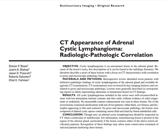
- “Though rare, an adrenal cystic lymphangioma should be suspected on CT when a unilocular or multilocular, low-attenuation, nonenhancing lesion is present in the region of the adrenal gland, particularly if the lesion contains either of the above-noted calcification patterns. Recognition of these findings may allow more conservative treatment of selected patients harboring these lesions.”
CT Appearance of Adrenal Cystic Lymphangioma: Radiologic-Pathologic Correlation
Steven P. Rowe, Justin A. Bishop, Jason D. Prescott, Roberto Salvatori, Elliot K. Fishman
AJR 2016; 206:81–85 - “ In all cases of adrenal lymphangioma from our institution, lesions appeared as well-circumscribed masses with overall low-attenuation internal contents (8– 20 HU on all contrast phases) . On contrast-enhanced images, all lesions had thin enhancing walls without evidence of nodularity. Though prior literature indicates that these lesions may have multiple thin septations, in our series two patients had at least one thin septation and the remaining patients had no septation visible on the CT examinations. The internal contents of the lymphangiomas did not otherwise quantitatively or visually enhance. Lesions varied in size between 2.5 and 6.0 cm (mean, 4.3 cm), and four had a maximum diameter greater than 4.0 cm.”
CT Appearance of Adrenal Cystic Lymphangioma: Radiologic-Pathologic Correlation
Steven P. Rowe, Justin A. Bishop, Jason D. Prescott, Roberto Salvatori, Elliot K. Fishman
AJR 2016; 206:81–85 - “Six of the seven adrenal lymphangiomas included in this report contained calcifications. The calcifications we encountered generally appeared in one of two patterns: thick, curvilinear, dystrophic-appearing calcifications or punctate and scattered peripheral calcifications.”
CT Appearance of Adrenal Cystic Lymphangioma: Radiologic-Pathologic Correlation
Steven P. Rowe, Justin A. Bishop, Jason D. Prescott, Roberto Salvatori, Elliot K. Fishman
AJR 2016; 206:81–85 - Purpose To investigate the proportion of clinically significant adrenal lesions in patients with a subcentimeter adrenal lesion, and the sensitivity of a cutoff size of 10 mm on computed tomography (CT).
Conclusion A lesion LD of ≥ 10 mm was a reasonable cutoff for determining adrenal abnormality. Subcentimeter lesions without visually high suspicion had a low risk of clinical significant lesions in our study cohort. Higher cutoffs significantly decreased sensitivity.
Clinical significance of a 10‐mm cutoff size for adrenal lesions: a retrospective study with 547 non‐oncologic patients undergoing adrenal computed tomography
Myoung Kyoung Kim et al.
Abdominal Radiology (2022) 47:1091–1097
- “Though rare, an adrenal cystic lymphangioma should be suspected on CT when a unilocular or multilocular, low-attenuation, nonenhancing lesion is present in the region of the adrenal gland, particularly if the lesion contains either of the above-noted calcification patterns. Recognition of these findings may allow more conservative treatment of selected patients harboring these lesions.”
CT Appearance of Adrenal Cystic Lymphangioma: Radiologic-Pathologic Correlation Steven P. Rowe, Justin A. Bishop, Jason D. Prescott, Roberto Salvatori, Elliot K. Fishman AJR 2016; 206:81–85 - “In all cases of adrenal lymphangioma from our institution, lesions appeared as well-circumscribed masses with overall low-attenuation internal contents (8– 20 HU on all contrast phases) . On contrast-enhanced images, all lesions had thin enhancing walls without evidence of nodularity. Though prior literature indicates that these lesions may have multiple thin septations, in our series two patients had at least one thin septation and the remaining patients had no septation visible on the CT examinations. The internal contents of the lymphangiomas did not otherwise quantitatively or visually enhance. Lesions varied in size between 2.5 and 6.0 cm (mean, 4.3 cm), and four had a maximum diameter greater than 4.0 cm.”
CT Appearance of Adrenal Cystic Lymphangioma: Radiologic-Pathologic Correlation Steven P. Rowe, Justin A. Bishop, Jason D. Prescott, Roberto Salvatori, Elliot K. Fishman AJR 2016; 206:81–85 - “ In all cases of adrenal lymphangioma from our institution, lesions appeared as well-circumscribed masses with overall low-attenuation internal contents (8– 20 HU on all contrast phases) . On contrast-enhanced images, all lesions had thin enhancing walls without evidence of nodularity. Though prior literature indicates that these lesions may have multiple thin septations, in our series two patients had at least one thin septation and the remaining patients had no septation visible on the CT examinations. The internal contents of the lymphangiomas did not otherwise quantitatively or visually enhance. Lesions varied in size between 2.5 and 6.0 cm (mean, 4.3 cm), and four had a maximum diameter greater than 4.0 cm.”
CT Appearance of Adrenal Cystic Lymphangioma: Radiologic-Pathologic Correlation Steven P. Rowe, Justin A. Bishop, Jason D. Prescott, Roberto Salvatori, Elliot K. Fishman AJR 2016; 206:81–85 - “Six of the seven adrenal lymphangiomas included in this report contained calcifications. The calcifications we encountered generally appeared in one of two patterns: thick, curvilinear, dystrophic-appearing calcifictions or punctate and scattered peripheral calcifications.”
CT Appearance of Adrenal Cystic Lymphangioma: Radiologic-Pathologic Correlation Steven P. Rowe, Justin A. Bish
- “Though rare, an adrenal cystic lymphangioma can be suspected on CT when a uni-locular or multi-locular, low-attenuation, non-enhancing lesion is present in the region of the adrenal gland, particularly if the lesion contains either of the above-noted calcification patterns. Recognition of these findings may allow more conservative treatment of selected patients harboring these lesions.”
MDCT Appearance of Adrenal Cystic Lymphangioma: Radiologic-Pathologic Correlation Steven P. Rowe , Justin A. Bishop , Jason D. Prescott , Roberto Salvatori, Elliot K. Fishman AJR (in press) - “Cystic lesions of the adrenal glands are among these rare tumors. In a previously reported series, most adrenal cystic lesions were pseudocysts (some of which were associated with other adrenal neoplasms), though endothelial cysts and an epithelial cyst were also reported. Other investigators have suggested that endothelial cysts are in fact more common than pseudocysts. Based on the types of lining cells in the cyst wall, adrenal endothelial cysts can be further subcategorized into angiomatous and lymphangiomatous cysts/cystic lymphangiomas. Cystic lymphangiomas of the adrenal gland have been estimated to be present in only 0.06% of the population.”
MDCT Appearance of Adrenal Cystic Lymphangioma: Radiologic-Pathologic Correlation Steven P. Rowe , Justin A. Bishop , Jason D. Prescott , Roberto Salvatori, Elliot K. Fishman AJR (in press) - “Further, calcifications in adenomas are unusual, generally occurring in large and degenerated adenomas . Calcification would certainly be more typical in old adrenal hematomas, and indeed the CT findings of adrenal lymphangioma are difficult to reliably distinguish from the sequela of prior hemorrhage given the low attenuation, non-enhancing nature of old hematomas. In any discussion of an adrenal mass with calcifications, the specter of adrenalcortical carcinoma can be raised, though again the lack of internal enhancement and the overall homogeneity of the lymphangiomas we have oberserved would generally not be seen in the case of this more ominous diagnosis.”
MDCT Appearance of Adrenal Cystic Lymphangioma: Radiologic-Pathologic Correlation Steven P. Rowe , Justin A. Bishop , Jason D. Prescott , Roberto Salvatori, Elliot K. Fishman AJR (in press)
- “Calcification would certainly be more typical in old adrenal hematomas, and indeed the CT findings of adrenal lymphangioma are difficult to distinguish from the sequelae of prior hemorrhage, given the shared characteristics of low attenuation, and non-enhancing nature. In any discussion of an adrenal mass with calcifications, the specter of adrenocortical carcinoma can be raised , though again the lack of internal enhancement and the overall homogeneity of the lymphangiomas we have oberserved would generally not be seen in the case of this more ominous diagnosis.”
MDCT Appearance of Adrenal Cystic Lymphangioma: Radiologic-Pathologic Correlation
Rowe SP, Bishop JA, Prescott JD, Salvatori R, Fishman EK
AJR (in press) - “The low attenuation on non-contrast CT can cause these lesions to be confused with lipid-rich adrenal adenomas, though the complete lack of internal enhancement on post-contrast images would not be consistent with the typical enhancement pattern seen with adenomas which generally enhance prominently and then wash out rapidly.Further, calcifications in adenomas are unusual, generally occurring in large and degenerated adenomas.”
MDCT Appearance of Adrenal Cystic Lymphangioma: Radiologic-Pathologic Correlation
Rowe SP, Bishop JA, Prescott JD, Salvatori R, Fishman EK
AJR (in press) - “The radiologic differential diagnosis of these lesions is varied and merits discussion. Interestingly, but perhaps not surprisingly given the rarity of this lesion, multiple reports have come to light in which an adrenal lymphangioma has been misidentified as arising from an adjacent organ. As might be predicted from the anatomic location of the adrenal gland, these reports have included cases in which an adrenal lymphangioma was thought to be a pancreatic tail cyst or a cystic lesion of the kidney.”
MDCT Appearance of Adrenal Cystic Lymphangioma: Radiologic-Pathologic Correlation
Rowe SP, Bishop JA, Prescott JD, Salvatori R, Fishman EK
AJR (in press) - “Cystic lesions of the adrenal glands are among these rare tumors. In a previously reported series, most adrenal cystic lesions were pseudocysts (some of which were associated with other adrenal neoplasms), though endothelial cysts and an epithelial cyst were also reported. Other investigators have suggested that endothelial cysts are in fact more common than pseudocysts. Based on the types of lining cells in the cyst wall, adrenal endothelial cysts can be further subcategorized into angiomatous and lymphangiomatous cysts/cystic lymphangiomas. Cystic lymphangiomas of the adrenal gland have been estimated to be present in only 0.06% of the population.”
MDCT Appearance of Adrenal Cystic Lymphangioma: Radiologic-Pathologic Correlation
Rowe SP, Bishop JA, Prescott JD, Salvatori R, Fishman EK
AJR (in press)
- “ Adrenal lymphangiomas, also known as cystic adrenal lymphangiomas, are rare, benign vascular lesions that usually remain asymptomatic throughout life. Although previously adrenal lymphangioma lesions were primarily found at autopsy, they are currently detected during imaging work-up for unrelated causes and are likely to imitate other adrenocortical or adrenal medullary neoplasms.”
Adrenal lymphangioma: clinicopathologic and immunohistochemical characteristics of a rare lesion.
Ellis CL et al
Human Pathol 2011 Jul;42(7)1013-8 - “Adrenal lymphangiomas are very rare, benign lymphatic neoplasms with a female, right-sided predominance in our current series. They may clinically present with abdominal pain or can be incidentally found during adulthood as a mass, necessitating surgical removal to rule out other types of adrenal neoplasms.”
Adrenal lymphangioma: clinicopathologic and immunohistochemical characteristics of a rare lesion.
Ellis CL et al
Human Pathol 2011 Jul;42(7)1013-8
