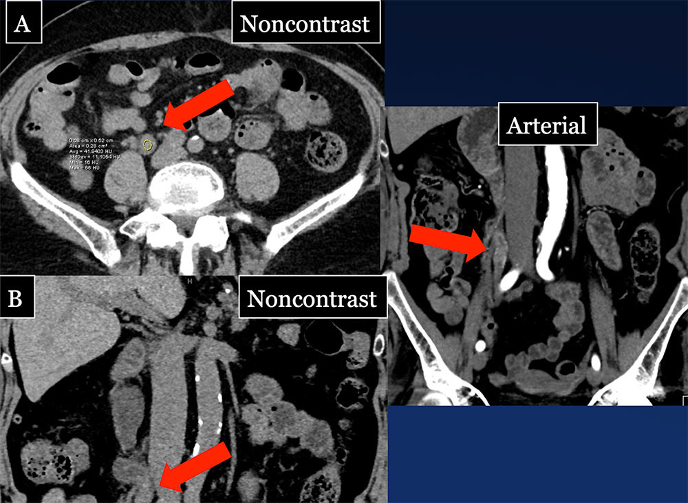
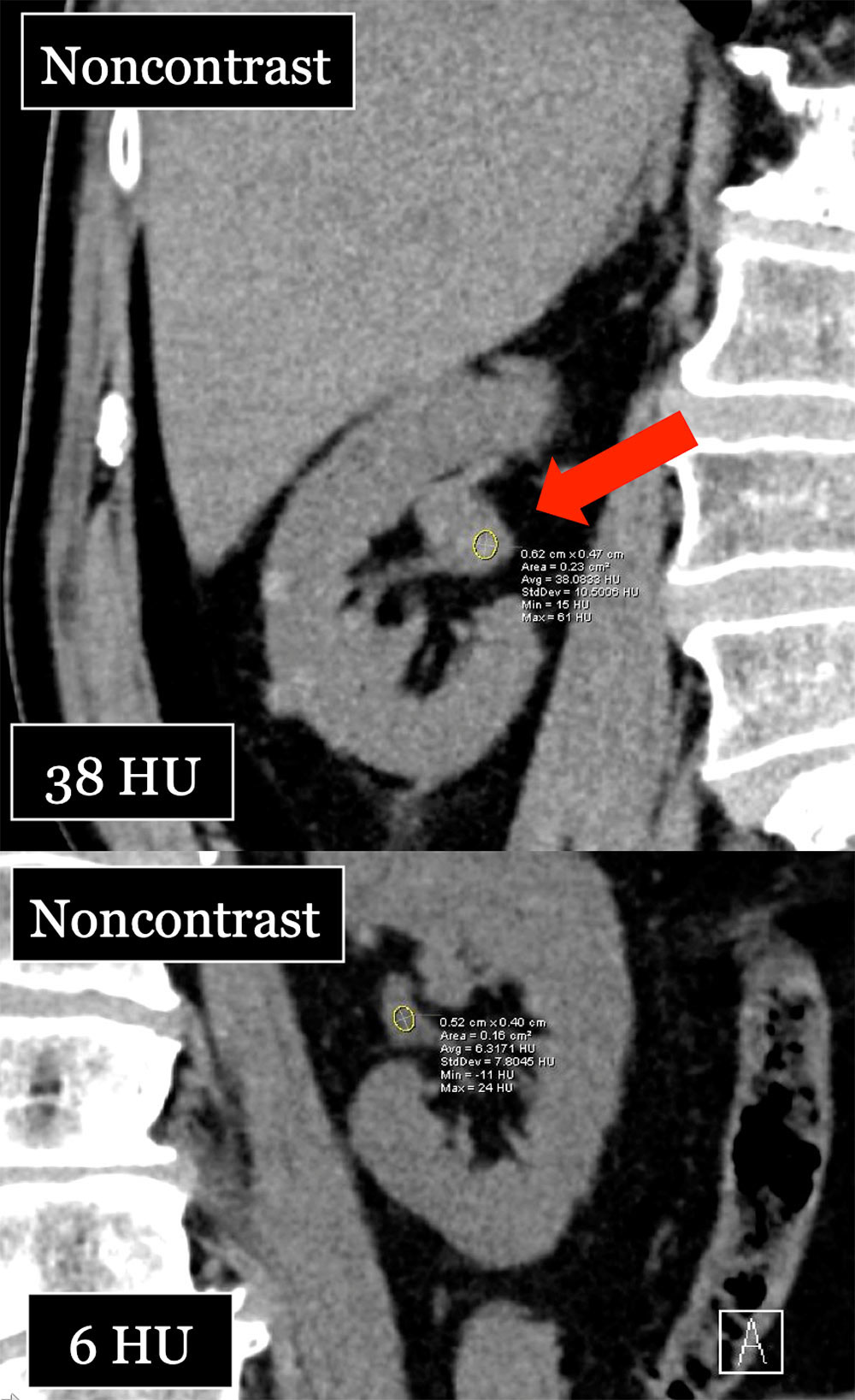
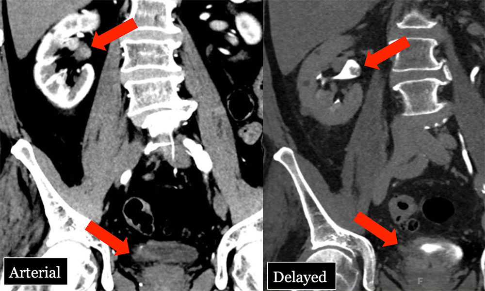
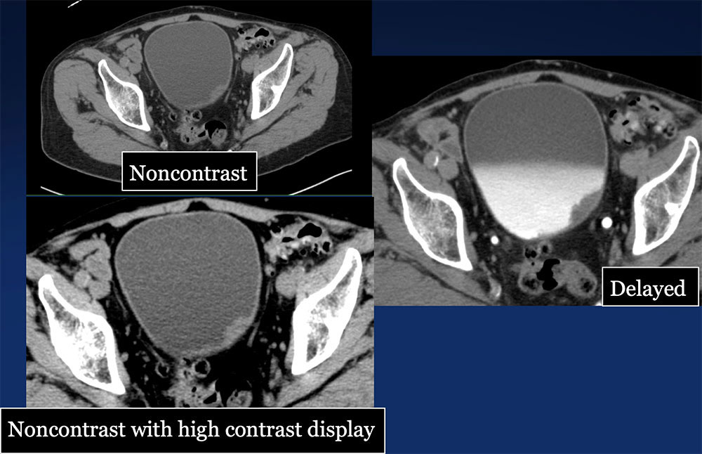
- Lipkin ME, Preminger GM. Imaging techniques for stone disease and methods for reducing radiation exposure. Urol Clin North Am. 2013 Feb;40(1):47-57.
- Johnson PT, Horton KM, Fishman EK. Noncontrast multidetector CT of the kidneys: utility of 2D MPR and 3D rendering to elucidate genitourinary pathology. Emerg Radiol. 2010 Jul;17(4):329-33.
- Vikram R, Sandler CM, Ng CS. Imaging and staging of transitional cell carcinoma: part 1, lower urinary tract. AJR 2009 Jun;192(6):1481-7.
- Vikram R, Sandler CM, Ng CS. Imaging and staging of transitional cell carcinoma: part 2, upper urinary tract. AJR 2009 Jun;192(6):1488-93.
- Westphalen AC, Hsia RY, Maselli JH, Wang R, Gonzales R. Radiological imaging of patients with suspected urinary tract stones: national trends, diagnoses, and predictors. Acad Emerg Med. 2011 Jul;18(7):699-707.
- Zwank MD1, Ho BM, Gresback D, Stuck LH, Salzman JG, Woster WR. Does computed tomographic scan affect diagnosis and management of patients with suspected renal colic? Am J Emerg Med. 2014 Apr;32(4):367-70.
- Moore CL, Daniels B, Singh D, Luty S, Molinaro A. Prevalence and clinical importance of alternative causes of symptoms using a renal colic computed tomography protocol in patients with flank or back pain and absence of pyuria. Acad Emerg Med. 2013 May;20(5):470-8.
- Pernet J, Abergel S, Parra J, Ayed A, Bokobza J, Renard-Penna R, Tostivint I, Bitker MO, Riou B, Freund Y. Prevalence of alternative diagnoses in patients with suspected uncomplicated renal colic undergoing computed tomography: a prospective study. CJEM. 2014 Feb 1;16(0):1-7.
- Katz DS, Scheer M, Lumerman JH, Mellinger BC, Stillman CA, Lane MJ. Alternative or additional diagnoses on unenhanced helical computed tomography for suspected renal colic: experience with 1000 consecutive examinations. Urology. 2000 Jul;56(1):53-7.
- Sountoulides P, Zachos I, Paschalidis K et al. Massive hematuria due to a congenital renal arteriovenous malformation mimicking a renal pelvis tumor: a case report. J Med Case Reports 2008; 2:144.
- Szolar DH, Kammerhuber F, Altziebler S, Tillich M, Breinl E, Fotter R, Schreyer HH. Multiphasic helical CT of the kidney: increased conspicuity for detection and characterization of small (<3-cm) renal masses. Radiology 1997; 202:211-217.
- Ramakrishnan K, Scheid DC. Diagnosis and management of acute pyelonephritis in adults. Am Fam Physician 2005; 71:933-942.
- Wong WS, Moss AA, Federle MP, Cochran ST, London SS. Renal infarction: CT diagnosis and correlation between CT findings and etiologies. Radiology 1984; 150:201-205.
- Westphalen A, Yeh B, Qayyum A, Hari A, Coakley FV. Differential diagnosis of perinephric masses on CT and MRI. AJR Am J Roentgenol. 2004; 183(6):1697-702.
- Browne RF, Meehan CP, Colville J, Power R, Torreggiani WC. Transitional cell carcinoma of the upper urinary tract: spectrum of imaging findings. Radiographics. 2005 Nov-Dec;25(6):1609-27.
- Ploeg M, Aben KK, Kiemeney LA. The present and future burden of urinary bladder cancer in the world. World J Urol 2009; 27:289.
- Jemal A, Bray F, Center MM, et al. Global cancer statistics. CA Cancer J Clin 2011; 61:69.
- Siegel R, Ma J, Zou Z, Jemal A. Cancer statistics, 2014. CA Cancer J Clin 2014; 64:9.
- Feldman AR, Kessler L, Myers MH, Naughton MD. The prevalence of cancer. Estimates based on the Connecticut Tumor Registry. N Engl J Med 1986; 315:1394.
- Babjuk M, Burger M, Zigeuner R, et al. EAU guidelines on non-muscle-invasive urothelial carcinoma of the bladder: update 2013. Eur Urol 2013; 64:639.
- Hall MC, Chang SS, Dalbagni G, et al. Guideline for the management of nonmuscle invasive bladder cancer (stages Ta, T1, and Tis): 2007 update. J Urol 2007; 178:2314.
- Babjuk M, Oosterlinck W, Sylvester R, et al. EAU guidelines on non-muscle-invasive urothelial carcinoma of the bladder. Eur Urol 2008; 54:303.
- Melicow MM. Tumors of urinary drainage tract: urothelial tumors. J Urol 1945; 54:186.
- Herr HW, Cookson MS, Soloway SM. Upper tract tumors in patients with primary bladder cancer followed for 15 years. J Urol 1996; 156:1286.
- Wallace DM, Raghavan D, Kelly KA, et al. Neo-adjuvant (pre-emptive) cisplatin therapy in invasive transitional cell carcinoma of the bladder. Br J Urol 1991; 67:608.
- Mitra AP, Skinner EC, Schuckman AK, et al. Effect of gender on outcomes following radical cystectomy for urothelial carcinoma of the bladder: a critical analysis of 1,994 patients. Urol Oncol 2014; 32:52.e1.
- Marshall VF. Current clinical problems regarding bladder tumors. Cancer 1956; 9:543.
- Khadra MH, Pickard RS, Charlton M, et al. A prospective analysis of 1,930 patients with hematuria to evaluate current diagnostic practice. J Urol 2000; 163:524.
- Grossman HB, Messing E, Soloway M, et al. Detection of bladder cancer using a point-of-care proteomic assay. JAMA 2005; 293:810.
- Dyer RB, Chen MY, Zagoria RJ. Classic signs in uroradiology. Radiographics. 2004;24 Suppl 1 (suppl 1): S247-80.
- Guimaraes AR & Harisinghani MG. Imaging of transitional cell carcinoma. In Vogelzang N ed. Comprehensive Textbook of Genitourinary Oncology. New York, NY: Lippincott Williams & Wilkins, 2006.
- Cosentino M, Palou J, Gaya JM, et al. Upper urinary tract urothelial: location as a predictive factor for concomitant bladder carcinoma. World J Urol 2013;31:141–5.
|
