google ads

High value MDCT angiography of acute superior mesenteric artery pathology: What the emergency medicine physician and vascular interventionist need to know

High value MDCT angiography of acute superior mesenteric artery pathology: What the emergency medicine physician and vascular interventionist need to know

Elliot K. Fishman, MD, FACR

The Russell H. Morgan Department of Radiology and Radiological Science
The Johns Hopkins Medical Institutions
Baltimore, Maryland


Imaging technique: Role of multi-planar reconstructions
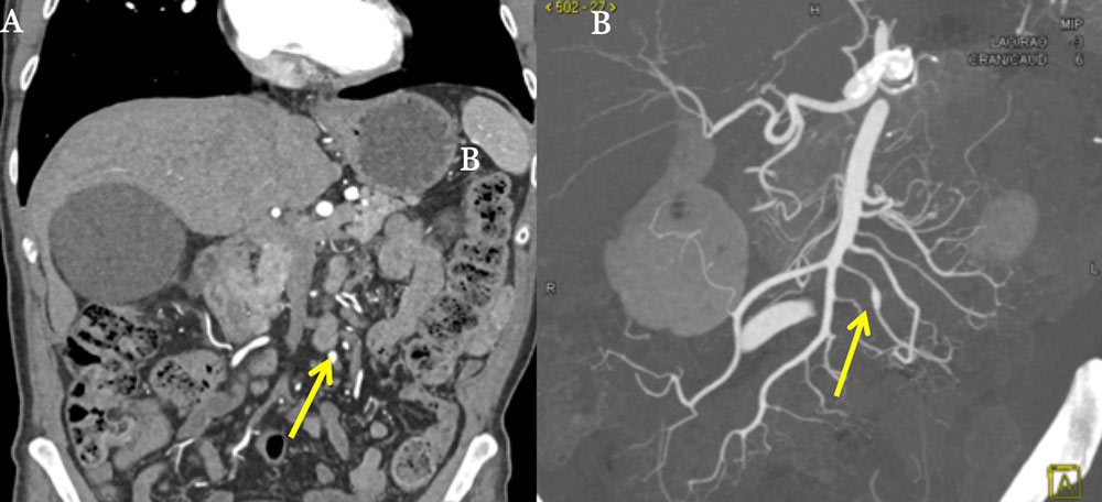
  • Standard coronal and sagittal reformats are generated at the scanner for each case
  • Real-time volumetric post-processing is performed at an independent workstation using 2D-multiplanar reformats (MPR), maximum Intensity Projection (MIP), 3D-volume rendering and 3D-cinematic rendering
  • Post-processing maximizes detection of small, subtle vascular lesions, and allows for evaluation of disease extent.
  • MIP using thin slabs allows visualization of the vasa recta.
74-year-old male with incidentally noted fusiform aneurysmal dilation of branch of SMA. It is difficult to identify the aneurysm on an MPR (A) (arrow), however, MIP (B) post-processing clearly elucidates the finding (arrow).
Imaging technique: Role of multi-planar reconstructions


Acute Mesenteric Ischemia (AMI)

  • Pathophysiology: abrupt decrease in either arterial or venous blood flow to bowel, with high morbidity and mortality (60-80%)
  • Causes: SMA embolus (40-50%), thrombus (~30%), mesenteric venous thrombus & non-occlusive mesenteric ischemia.
    • Emboli from the heart lodge in the SMA a few centimeters distal to the origin, near the origin of the middle colic artery. Smaller emboli lodge in distal branches.
    • Thrombosis resulting from rupture of an unstable atherosclerotic plaque, more likely to occur in the proximal 2 cm of the SMA.
    • Non-occlusive ischemia occurs in the setting of hypotension or cardiogenic shock with hypoperfusion resulting in vasoconstriction.
  • Clinical presentation: severe abdominal pain, nausea, vomiting, and diarrhea
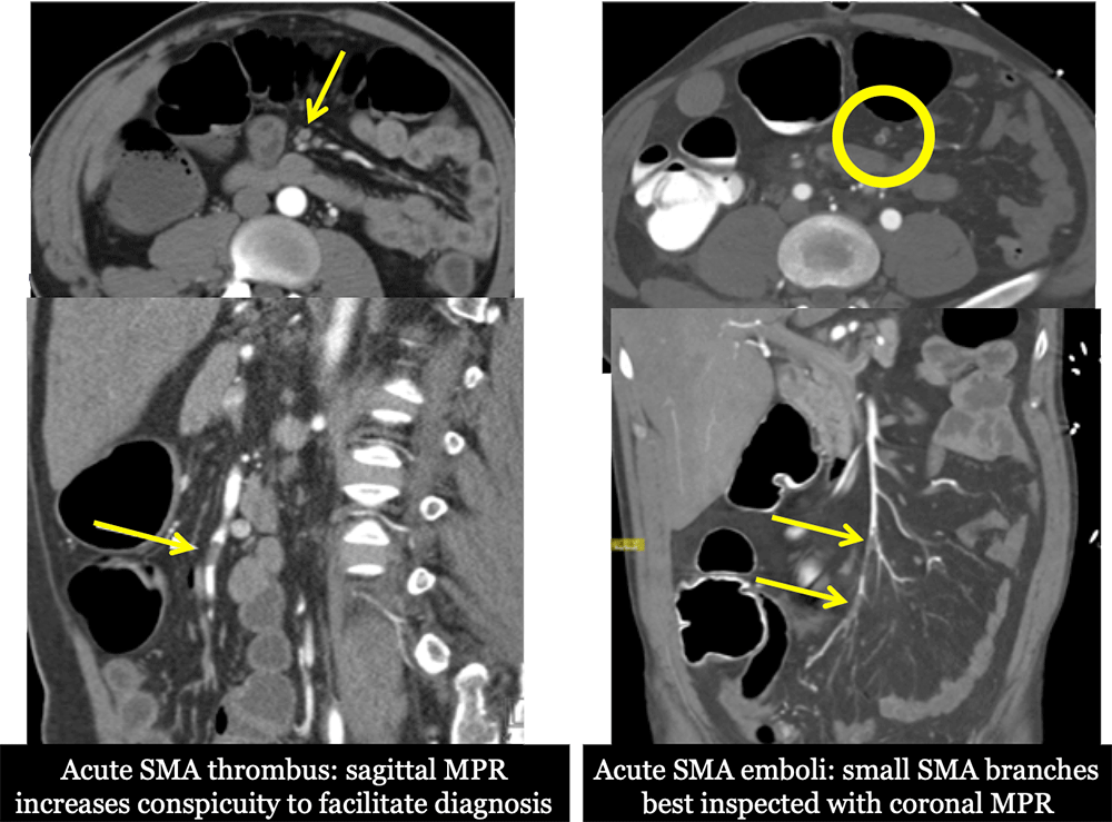
  • In the setting of acute thrombus, CT demonstrates a low density filling defect or vessel cutoff sign, best seen on sagittal view.
  • Coronal 2D and 3D renderings facilitate visualization of distal branch occlusions.


Acute Mesenteric Ischemia (AMI)

Illustration demonstrating thrombosis of the SMA with sharp vessel cut-off.
Acute Mesenteric Ischemia (AMI)


Acute Mesenteric Ischemia (AMI): MPRs

Acute Mesenteric Ischemia (AMI): MPRs


Acute Mesenteric Thrombosis

82-year-old male with a history of HTN, sick sinus syndrome and idiopathic cardiomyopathy who presented with severe abdominal pain, diarrhea, and emesis, elevated lactate level and acute kidney injury. Coronal (A), sagittal (B), and MIP (C) CT images demonstrate thrombosis and complete occlusion of the distal SMA with a sharp cutoff (red arrow).

Acute Mesenteric Thrombosis


Chronic Mesenteric Ischemia (CMI)

  • Leading cause is atherosclerosis (95%).
  • Other causes include median arcuate ligament syndrome, arterial dissection, FMD, and radiation.
  • Mortality is approximately 40-50%
  • Recurrent episodes of postprandial abdominal pain referred to as intestinal angina.
  • Weight loss due to food aversion, nausea, vomiting, and diarrhea.
  • Usually only become symptomatic when 2 of 3 major mesenteric vessels are severely stenotic or occluded, although stenosis of 1 major vessel can produce symptoms.
  • MDCT findings: SMA wall thickening, calcified plaque, and/or filling defects caused by non-calcified plaque.
  • Most common in the proximal 2 cm of SMA near the ostium.
  • Collateral vessels may be present to compensate.
Acute Mesenteric Thrombosis

Illustration demonstrating atherosclerotic plaque narrowing the proximal 2 cm of SMA near the ostium


Chronic SMA Thrombosis

56-year-old male with a history of HTN, HLD, CAD, and COPD who presents with severe postprandial abdominal pain. He has started to avoid meals and lost 10 pounds in the past month. Coronal (A), sagittal (B), MIP (C), and volume rendering (D) CT images demonstrate an approximately 2 cm segment of occlusive thrombus within the proximal SMA. The SMA reconstitutes distally by several collateral vessels.

Chronic SMA Thrombosis


SMA Dissection

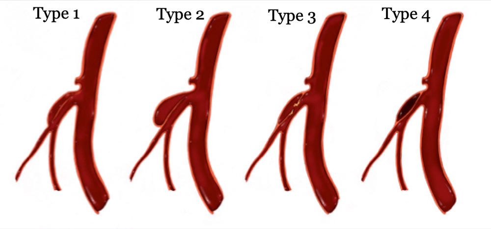
  • Several different classification schemes exist for isolated SMA dissections. Sakamoto et al. describe four types:
    • Type 1 has a patent false lumen with entry and re-entry.
    • Type 2 is a “cul-de-sac” shaped false lumen without re-entry.
    • Type 3 has a thrombosed false lumen with ulcer.
    • Type 4 has a completely thrombosed false lumen without ulcer.
  • Treatment for the different types has not been adequately evaluated.
SMA Dissection


SMA Dissection

48-year-old female who presents with diffuse abdominal pain and tearing back pain associated with nausea and vomiting. Axial (A) and sagittal (B) CT images demonstrate a short segment nonocclusive type II dissection of the mid SMA (red arrows) without bowel wall thickening or ischemia, amenable to stenting. The 3D volume rendering sagittal (C) and coronal (D) CT images depict the extent of the dissection (yellow arrows).

SMA Dissection


SMA Dissection

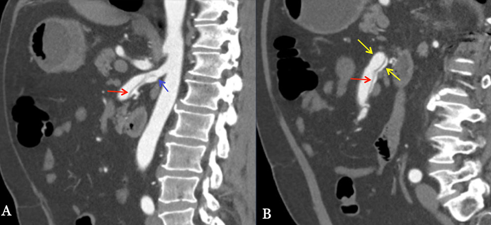
56-year-old male with Marfan syndrome. Sagittal images (A and B) demonstrating the long segment of SMA dissection with the flap extending 11 cm. An aneurysm of the mid-SMA measuring 1.7 cm is identified by the yellow arrows. The false lumen is patent and provides flow to multiple intestinal branches. The start of the dissection flap (blue arrow) is 1.4 cm from the origin of the SMA. All findings are important to know for possible endovascular treatment.
SMA Dissection


SMA Dissection

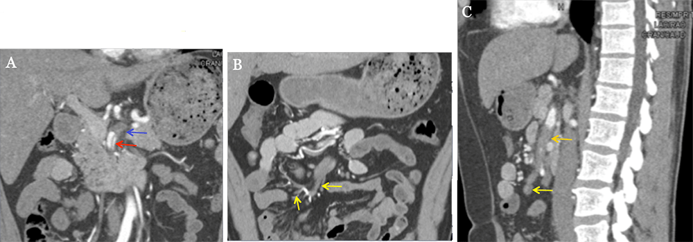
49-year-old male who presents with acute onset abdominal pain radiating to his back pain. Coronal (A and B) and sagittal (C) CT images demonstrate a proximal dissection of the SMA (red arrow) extending into jejunal and ileal branches (yellow arrows). Thrombosed and dilated proximal false lumen depicted by the blue arrow. Near complete occlusion of the distal SMA (orange arrow).
SMA Dissection


SMA Dissection

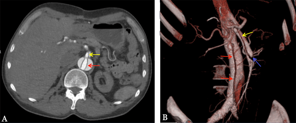
59-year-old male with a type B aortic dissection. Axial image (A) and the volume rendering 3D image demonstrate the descending aorta dissection flap (red arrow) involving the SMA (yellow arrow), precluding primary repair with endovascular stenting. The volume rendering 3D image depicts the blind ending false lumen with intestinal branches (blue arrow).
SMA Dissection


SMA Aneurysm

29-year-old male with a history of Loeys-Dietz syndrome who presents with abdominal pain, nausea, and vomiting. Axial (A), sagittal (B), and 3D rendering (C) CT images demonstrate fusiform aneursymal dilation of the proximal SMA measuring up to 3.5 cm (red arrows) with dilation of the more distal SMA to 1.2 cm (yellow arrows). Minimal wall thickening and fat stranding surrounding the proximal SMA. Tortuosity and dilation of the abdominal aorta and common iliac arteries.

SMA Aneurysm


SMA Aneurysm

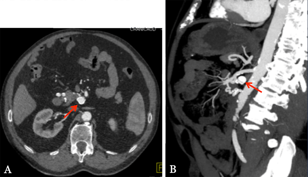
82-year-old male with proximal celiac artery occlusion who presents with peri-umbilical abdominal pain. Axial (A) image demonstrates a calcified enhancing lesion arising from an unclear vessel (red arrow). Multiple collateral vessels arising from the pancreaticoduodenal arcade. The sagittal MIP (B) image more clearly demonstrates a saccular aneurysm of a proximal SMA branch measuring up to 1.7 cm (red arrows).
SMA Aneurysm


SMA Aneurysm

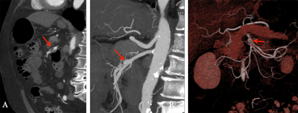
69-year-old male with abdominal pain and an incidental aneurysm found at outside hospital. Sagittal image (A) demonstrates a partially thrombosed aneurysm of the SMA (red arrow). The sagittal MIP image (B) easily depicts the aneurysm is 4.2 cm from the SMA origin. Volume rendering 3D image (C) shows the location of the aneurysm in relation to the branches of the SMA and the pancreas.
SMA Aneurysm


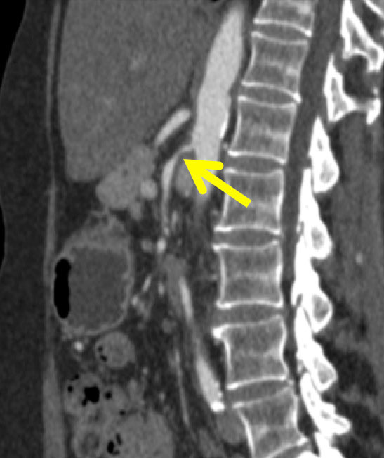
SMA Vasculitis

70-year-old female with a history of Takayasu’s arteritis who presents with abdominal pain. Sagittal MPR shows proximal circumferential narrowing (arrow). Endovascular treatment may be attempted if medical management fails.
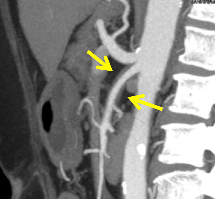
SMA Vasculitis
77-year-old female with a history of giant cell arteritis who presents with weight loss. Sagittal MIP image shows the long segment circumferential narrowing (arrow) without critical stenosis. Endovascular treatment is not advised for this patient.
SMA Vasculitis


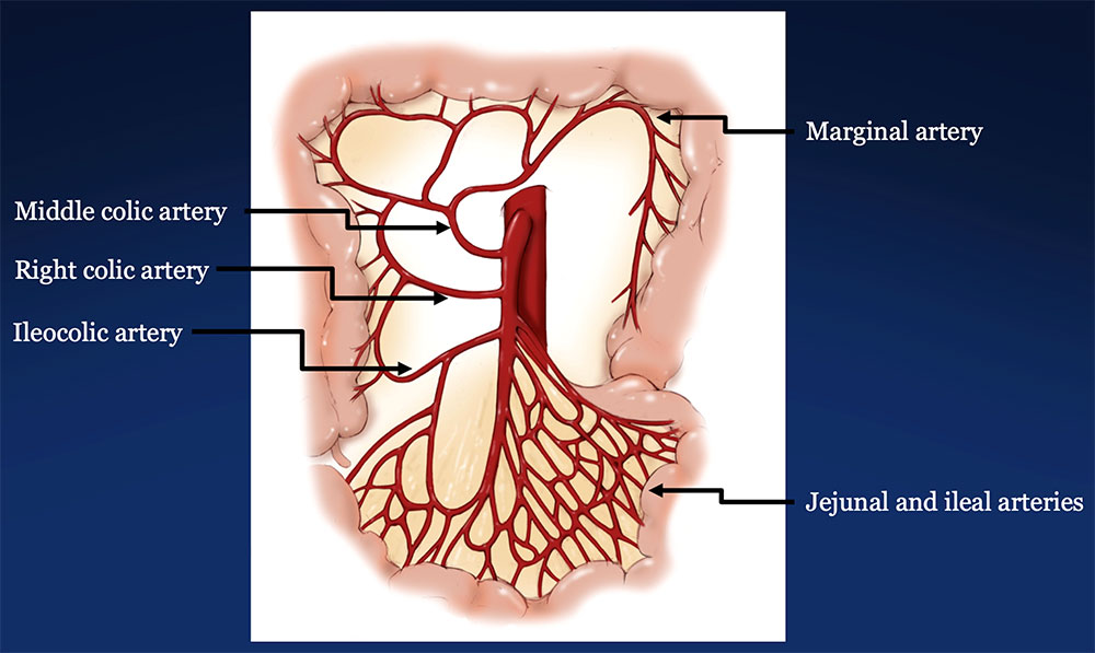
SMA Vascular Territory

Illustration demonstrating SMA vascular territory from near the ligament of treitz to the proximal two-thirds of the transverse colon.
SMA Vascular Territory


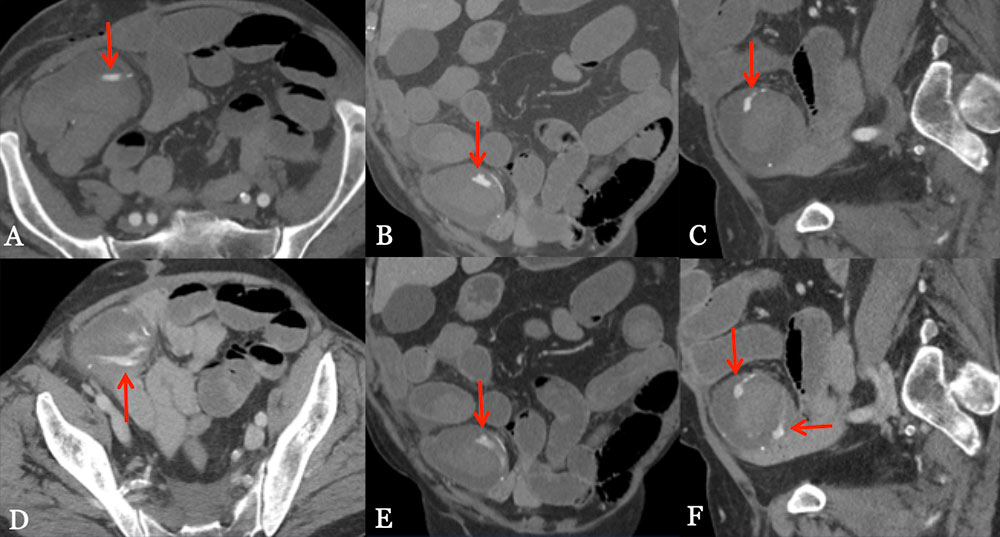
SMA Hemorrhage

76-year-old male with a history of rectal adenocarcinoma status post low anterior resection and recent diverting ileostomy takedown who presents with large volume bright red blood per rectum. Axial (A and D), coronal (B and E), and sagittal (C and F) CT images in the arterial (A-C) and venous (D-F) phase demonstrate contrast extravasation at the site of ileal anastomosis in the RLQ with pooling of blood.
SMA Hemorrhage


SMA Hemorrhage

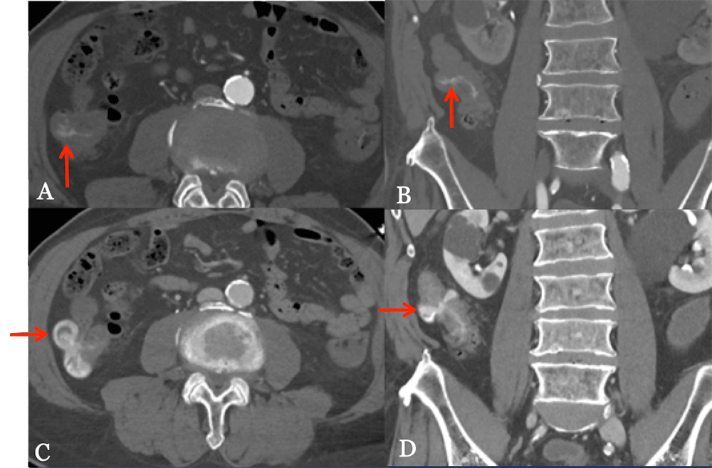
84-year-old male with a history of atrial fibrillation and sick sinus syndrome who presents with bright red blood per rectum. Axial (A and C) and coronal (B and D) CT images in the arterial (upper images) and venous (lower images) phase demonstrate hyperdense material in the ascending colon as well as within a diverticulum that increases on venous phase, compatible with a bleeding diverticulum (red arrows).
SMA Hemorrhage


Refererences

  • Angle FJ, Nida BA, and Matsumoto A. Managing mesenteric vasculitis. Techniques in Vascular and Interventional Radiology. 2015; 18(1): 38-42.
  • Belli AM, Markose G, and Morgan R. The role of interventional radiology in the management of abdominal visceral artery aneurysms. Cardiovascular and Interventional Radioloy. 2012; 35(2): 234-243.
  • Chen JK, Johnson PT, Horton KM, and Fishman EK. Unsuspected mesenteric arterial abnormality: comparison of MDCT axial sections to interactive 3D rendering. American Journal of Roentgenology. 2007; 189(4): 807-813.
  • Dong Z, Fu W, Chen B, et al. Treatment of symptomatic isolated dissection of superior mesenteric artery. Journal of Vascular Surgery. 2013; 57(2): 69s-76s.
  • Foley MI, Moneta GL, Abou-Zamzam AM, et al. Revascularization of the superior mesenteric artery alone for treatment of intestinal ischemia. Journal of Vascular Surgery. 2000; 31(1): 37-47.
  • Garrett Jr. HE. Options for treatment of spontaneous mesenteric artery dissection. Journal of Vascular Surgery. 2014; 59(5): 1433-1439.


Refererences

  • Ha HK, Lee SH, Rha SE, et al. Radiologic features of vasculitis involving the gastrointestinal tract. Radiographics. 2000; 20(3): 779-794.
  • Hagspiel KD, Flors L, Hanley M, and Norton PT. Computed tomography angiography and magnetic resonance angiography imaging of the mesenteric vasculature. Techniques in Vascular and Interventional Radiology. 2015; 18(1): 2-13.
  • Hemp JH and Sabri SS. Endovascular management of visceral arterial aneurysms. Techniques in Vascular and Interventional Radiology. 2015; 18(1): 14-23.
  • Horton KM and Fishman EK. CT angiography of the mesenteric circulation. Radiologic Clinics of North America. 2010; 48(2): 331-345.
  • Jia Z, Tu J, and Jiang G. The classification and management strategy of spontaneous isolated superior mesenteric artery dissection. Korean Circulation Journal. 2017; 47(4): 425-431.
  • Jiang J, Ding X, Su Q, et al. Therapeutic management of superior mesenteric artery aneurysms. Journal of Vascular Surgery. 2011; 53(6): 1619-1624.


Refererences

  • Laing CJ, Tobias T, Rosenblum DI, et al. Acute gastrointestinal bleeding: emerging role of multidetector CT angiography and review of current imaging techniques. Radiographics. 2007; 27(4): 1055-1070.
  • Luan JY, Guan X, Li X, et al. Isolated superior mesenteric artery dissection in China. Journal of Vascular Surgery. 2016; 63(2): 530-536.
  • Luan JY, Li X, Li TR, et al. Vasodilator and endovascular therapy for isolated superior mesenteric artery dissection. Journal of Vascular Surgery. 2013; 57(6): 1612-1620.
  • Matsumoto AH, Angle JF, Spinosa DJ, et al. Percutaneous transluminal angioplasty and stenting in the treatment of chronic mesenteric ischemia: results and longterm followup. Journal of the American College of Surgeons. 2002; 194(1): S22-S31.
  • Min S, Yoon KC, Min SK, et al. Current strategy for the treatment of symptomatic spontaneous isolated dissection of superior mesenteric artery. Journal of Vascular Surgery. 2011; 54(2): 461-466.
  • Oderich GS, Bower TC, Sullivan TM, et al. Open versus endovascular revascularization for chronic mesenteric ischemia: risk-stratified outcomes. Journal of Vascular Surgery. 2009; 49(6): 1472-1479.


Refererences

  • Rawat N and Gibbons CP. Surgical and endovascular treatment for chronic mesenteric ischemia: a multicenter study. Annals of Vascular Surgery. 2010; 24(7): 935-945.
  • Rits Y, Oderich GS, Bower TC, et al. Interventions for mesenteric vasculitis. Journal of Vascular Surgery. 2010; 51(2): 392-400.
  • Sakamoto I, Ogawa Y, Sueyoshi E, et al. Imaging appearances and management of isolated spontaneous dissection of the superior mesenteric artery. European Journal of Radiology. 2007; 64(1): 103-110.
  • Sasae Y, Morita Y, Sakuta T, et al. Abdominal pain as the initial presentation of Takayasu arteritis. Modern Rheumatology. 2008; 18(5): 496-498.
  • Stone WM, Abbas M, Cherry KJ, et al. Superior mesenteric artery aneurysms: is presence an indication for intervention? J Vasc Surg. 2002; 36: 234–237.
  • Verde F, Bleich KB, Oshmyansky A, et al. Isolated celiac and superior mesenteric artery dissection identified with MDCT: imaging findings and clinical course. Journal of Computer Assisted Tomography. 2012; 36: 539-545.


Refererences

  • Verma H, Oderich GS, and Tripathi RK. Surgical and endovascular interventions for chronic mesenteric ischemia. Journal of Cardiovascular Surgery. 2015; 56(2): 299-307.
  • Zhao YE, Wang ZJ, Zhou CS, Zhu FP, Zhang LJ, and Lu GM. Multidetector computed tomgraphy of superior mesenteric artery: anatomy and pathologies. Canadian Association of Radiologists Journal. 2014; 65(3): 267-274.

Privacy Policy

Copyright © 2025 The Johns Hopkins University, The Johns Hopkins Hospital, and The Johns Hopkins Health System Corporation. All rights reserved.