- 3
- 1
- 6
To Quiz Yourself: Select OFF by clicking the button to hide the diagnosis & additional resources under the case.
Quick Browser: Select ON by clicking the button to hide the additional resources for faster case review.
CASE NUMBER
116


Diagnosis
Osler-Weber-Rendu syndrome
Note
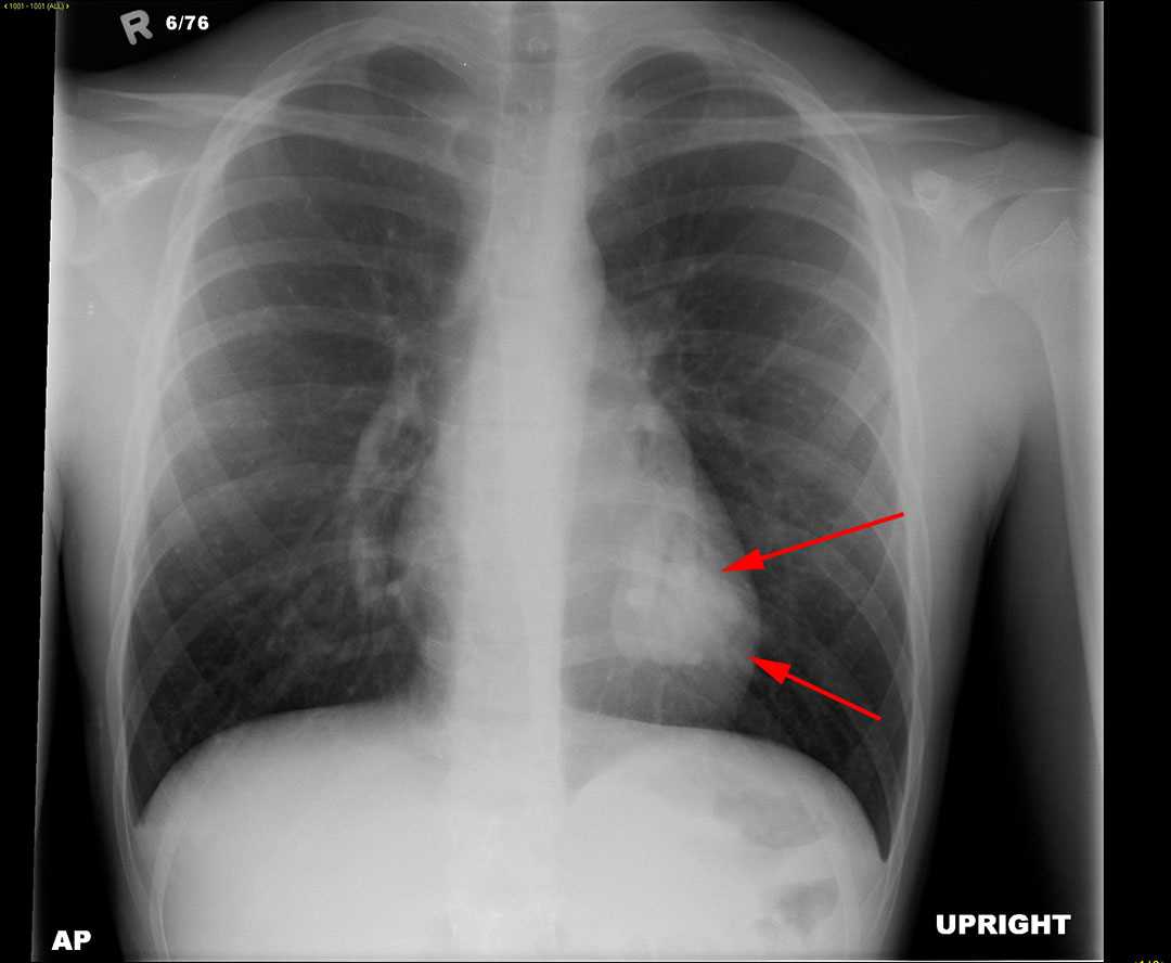
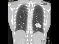
This is a patient who presents with recurrent nose bleeds and shortness of breath. A frontral radiograph of the chest was obtained which demonstrates a mass in the left lower lobe as indicated by the red arrows. To further evaluate this mass, a contrast enhanced CT of the chest was obtained. The second image is a maximum intensity projection reconstruction in the coronal plane in lung windows. The lobulated mass in the left lower lobe is similar in attenuation to the small surrounding pulmonary vessels and its morphology resembles a tangle of large vessels. The third image is a maximum intensity projection reconstruction of the chest in the coronal plane. The red arrow demonstrates the branch of the left lower lobe pulmonary artery that feeds the lesion. The green arrow indicates the dilated left lower lobe pulmonary vein that drains the lesion. The findings are most compatible with a large pulmonary arteriovenous malformation. Pulmonary arteriovenous malformations as well as recurrent nose bleeds are seen in a condition known as hereditary hemorrhagic telangiectasias, which is also known as Osler-Weber-Rendu syndrome. These patients acquire arteriovenous malformations throughout the body and brain. Pulmonary arteriovenous malformations are treated with coil embolization to prevent complications such as strokes as well as brain abscesses.
Related videos to the case

THIS IS CASE
116
OF
129