- 3
- 1
- 6
To Quiz Yourself: Select OFF by clicking the button to hide the diagnosis & additional resources under the case.
Quick Browser: Select ON by clicking the button to hide the additional resources for faster case review.
CASE NUMBER
82
Diagnosis
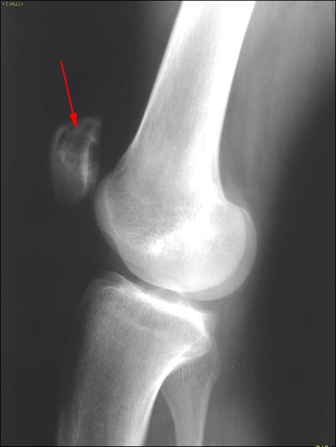
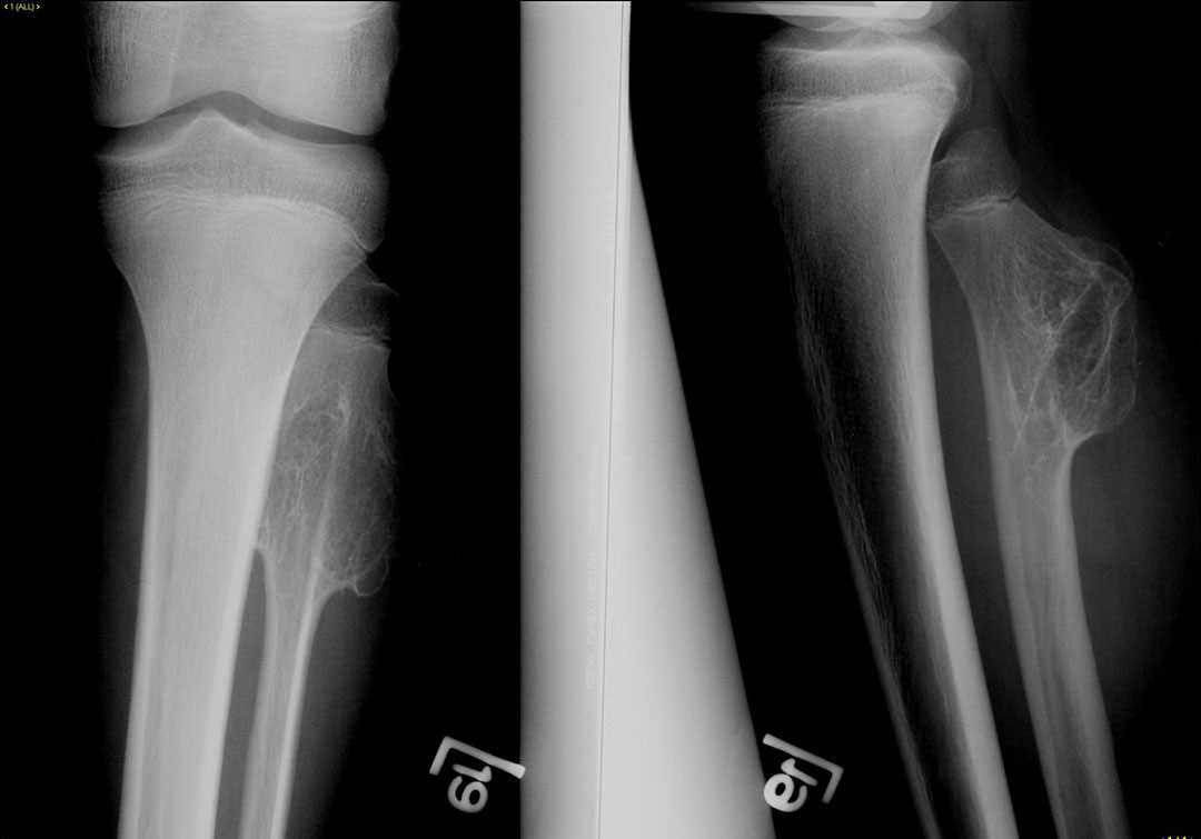
Osteochondroma of the fibula
Note
This is a case of a 13 year male who presented with firm swelling of the calf on physical examination. Radiographs of the tibia and fibula demonstrate an osseous mass arising from the posterior aspect of the fibula. The mass has wide-based origin from the fibula consistent with a sessile lesion. The mass looks like normal bone and both the medullary cavity and cortex of the fibula are contiguous with the lesion. Findings are most suggestive of an exostosis also known as an osteochondroma. Pain is a typical presentation of these tumors. Complications can include fracture, bursitis, or impingement on adjacent neurovascular bundle. Although benign, these tumors are monitored radiographically as there is a small risk of malignant transformation into chondrosarcoma. MRI can be used to assessed the cartilage cap overlying these tumors, as thicker cartilage caps are associated with a greater risk of malignant transformation.
Related videos to the case


THIS IS CASE
82
OF
129