- 3
- 1
- 6
To Quiz Yourself: Select OFF by clicking the button to hide the diagnosis & additional resources under the case.
Quick Browser: Select ON by clicking the button to hide the additional resources for faster case review.
CASE NUMBER
77


Diagnosis
Intraosseous lipoma
Note
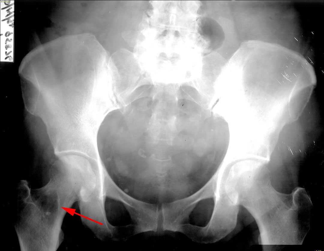
This a 31 year old male who presented with left hip pain. A frontal radiograph of the pelvis demonstrated a well circumscribed lesion with a narrow zone of transition in the intertrochanteric portion of the right femur. The central attenuation of the lesion is low suggestive of possible fat. There is a focal calcification within the lession. Image 2 is a CT of the right femur which demonstrates an intraosseous fat containing lesion with central calcification. The third image is a T1 weighted MR image of the right femur. The mass is centrally T1 hyperintense consistent with fat, similar in appearance to the surrounding normal fatty marrow. The lesion location and imaging characteristics are typical for an intraosseous lipoma. If this lesion was more heterogeneous, an additional fat containing lesion in this region such as a liposclerosing myxofibrous tumor would have to be considered.
Related videos to the case


THIS IS CASE
77
OF
129