- 2
- ,
- 2
- 4
- 6
To Quiz Yourself: Select OFF by clicking the button to hide the diagnosis & additional resources under the case.
Quick Browser: Select ON by clicking the button to hide the additional resources for faster case review.
CASE NUMBER
270


Diagnosis
Tuberous Sclerosis with Subependymal Giant Cell Astrocytoma
Note
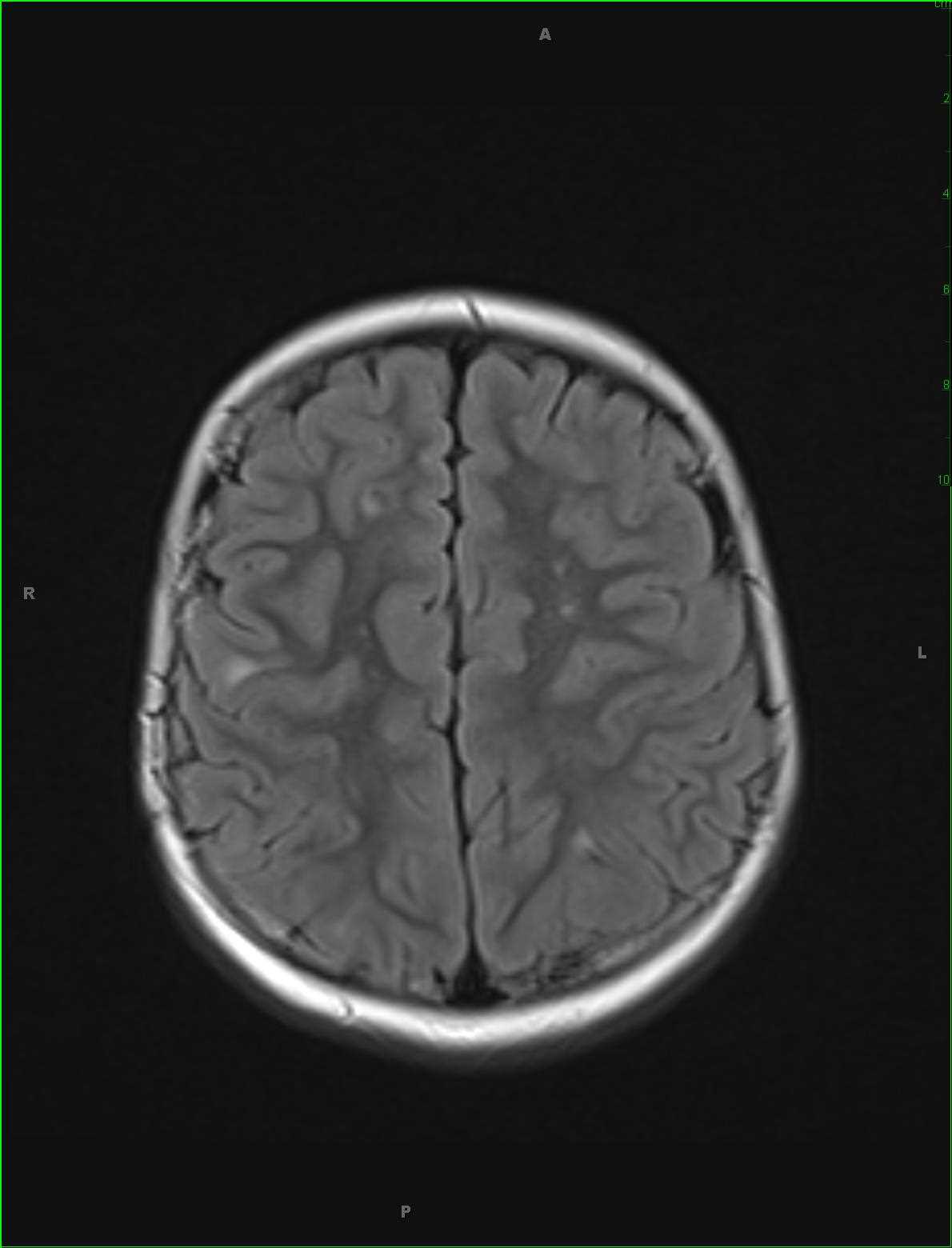
12-year-old male with history of seizures. The fluid sensitive images demonstrate nodular regions of hyperintense signal in the subcortical white matter of the supratentorial compartment, some of which is more linear and radiating towards the ventricular margins. On the anatomic T1 weighted images, there are regions of nodularity along the margins of the lateral ventricles. A prominent focus of susceptibility is identified within the anterior aspect of the corpus callosum body. There is an additional rounded focus of susceptibility at the anterior aspect of the sylvian fissure on the left on the T2 axial image. On the postcontrast images, there are enhancing ovoid lesions in the frontal horns of each lateral ventricle with the most prominent lesion central right. The imaging findings are compatible with tuberous sclerosis. Tuberous sclerosis falls in the category of phakomatosis or neuro-oculocutaneous syndromes. Clinical presentation typically includes seizures, mental retardation, and adenoma sebaceum. Tuberous sclerosis results from mutations in tumor suppressor genes, TSC1 which is on chromosome 9 and TSC2, chromosome 16.
Related videos to the case