- 2
- ,
- 2
- 5
- 2
To Quiz Yourself: Select OFF by clicking the button to hide the diagnosis & additional resources under the case.
Quick Browser: Select ON by clicking the button to hide the additional resources for faster case review.
CASE NUMBER
266


Diagnosis
Second Branchial Cleft Cyst, with Rupture
Note
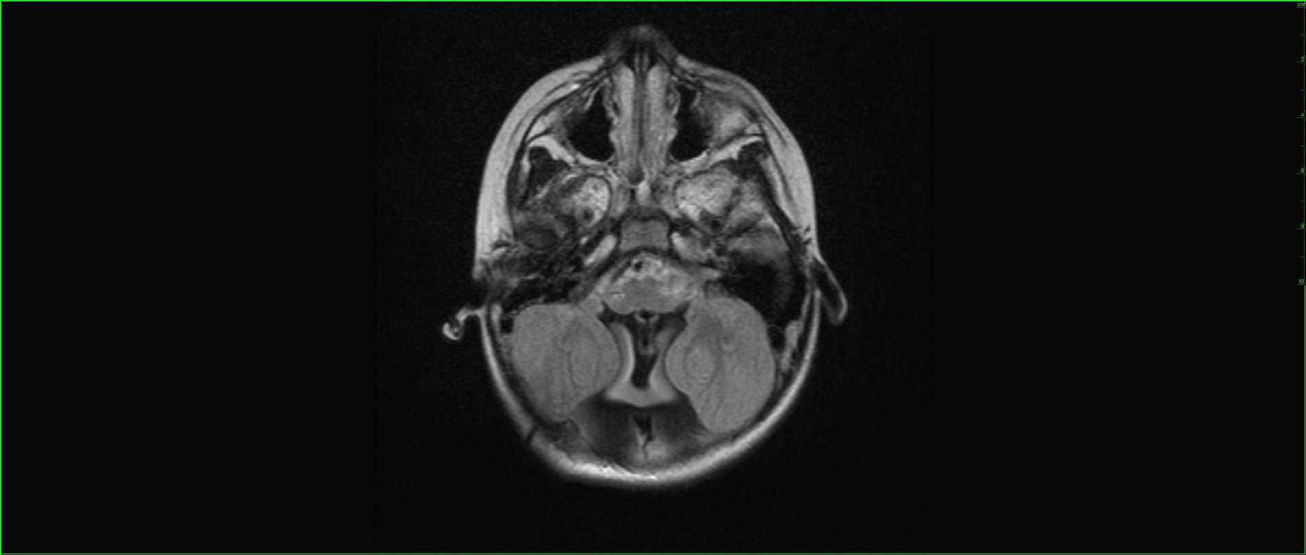
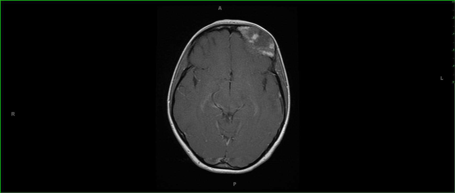
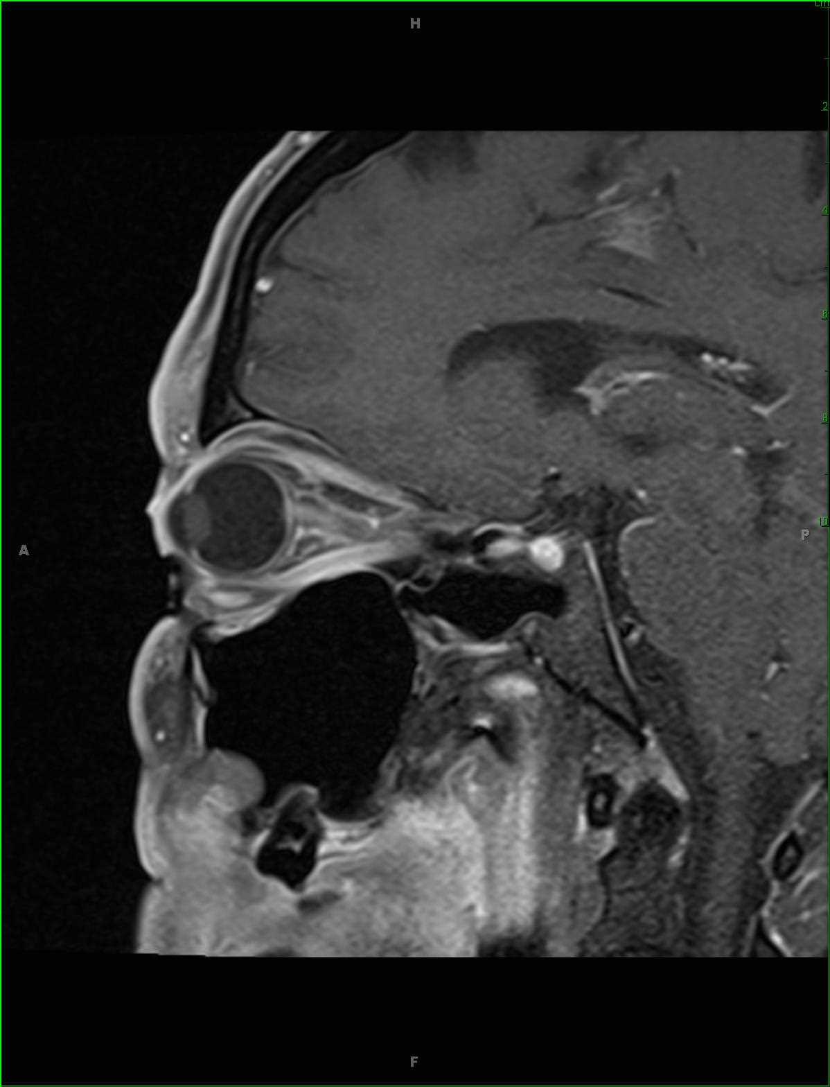
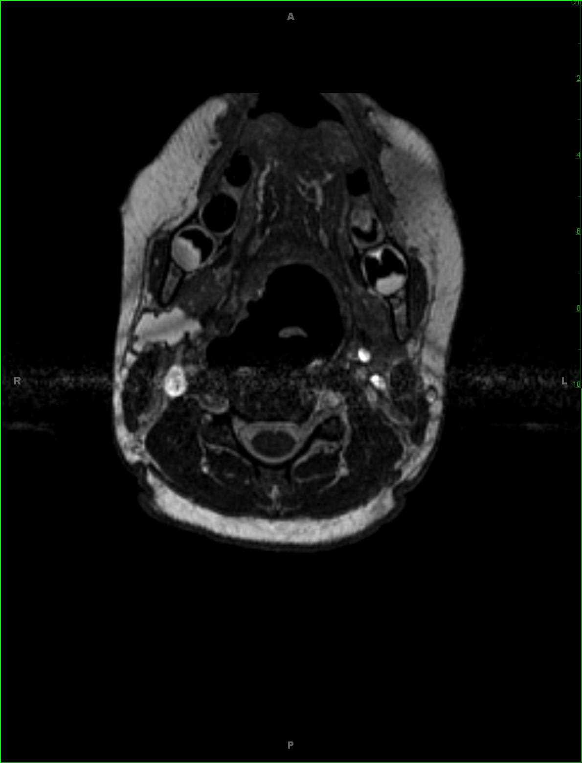
20-something-year-old male with recurrent swelling and pain at the right mandibular angle. There is a circumscribed T2 and proton density hyperintense lesion arising at the posterior margin of the mandibular ramus and extending posterior to the submandibular gland. The lesion demonstrates well defined borders with extralesional T2 and proton density hyperintense signal. The differential includes second branchial cleft cyst with rupture more likely than lymphatic malformation, dermoid, and lymph node with necrosis. On resection, this lesion was found to be a second branchial cleft cyst. Second branchial cleft cyst result from cystic dilatations of the remnant of the second branchial apparatus. Presentation in early adulthood is common, typically occurring after minor trauma or infection. Branchial cleft sinuses or fistula typically present earlier. Typical clinical features of a branchial cleft cyst include a rounded swelling just below the angle of the mandible anterior to the sternocleidomastoid muscle. Treatment is with complete surgical resection.
Related videos to the case