- 2
- ,
- 3
- 8
- 1
To Quiz Yourself: Select OFF by clicking the button to hide the diagnosis & additional resources under the case.
Quick Browser: Select ON by clicking the button to hide the additional resources for faster case review.
CASE NUMBER
245
Diagnosis
Chordoma
Note
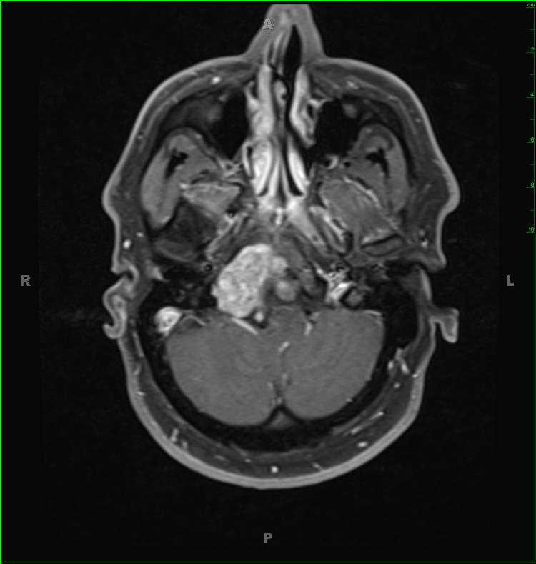
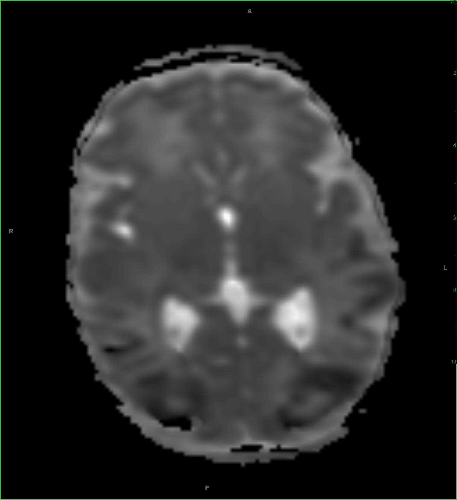
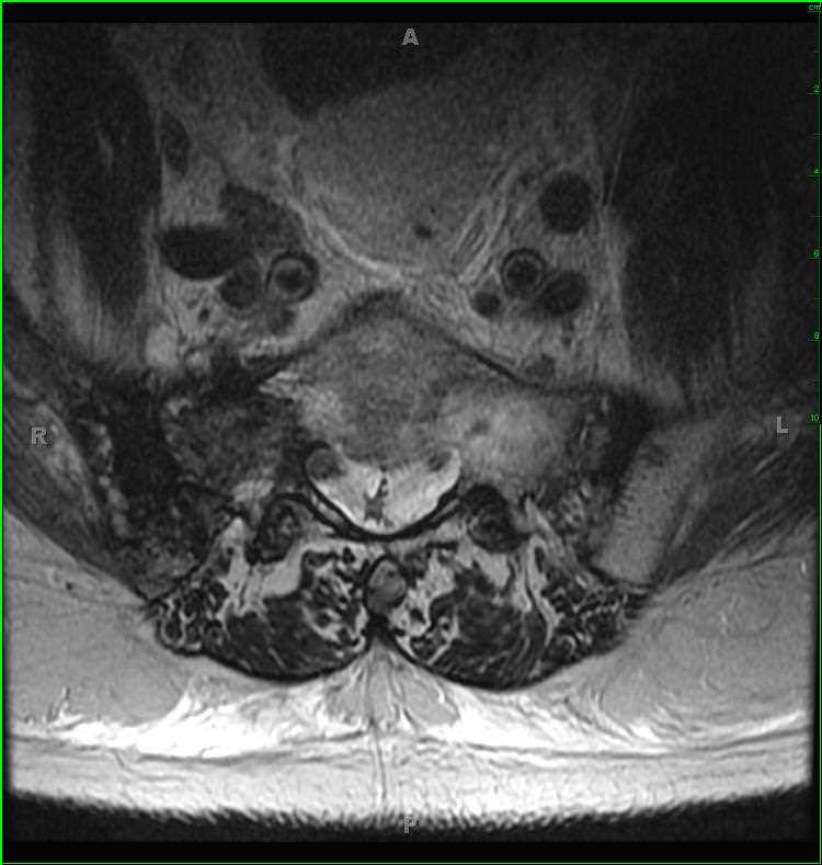
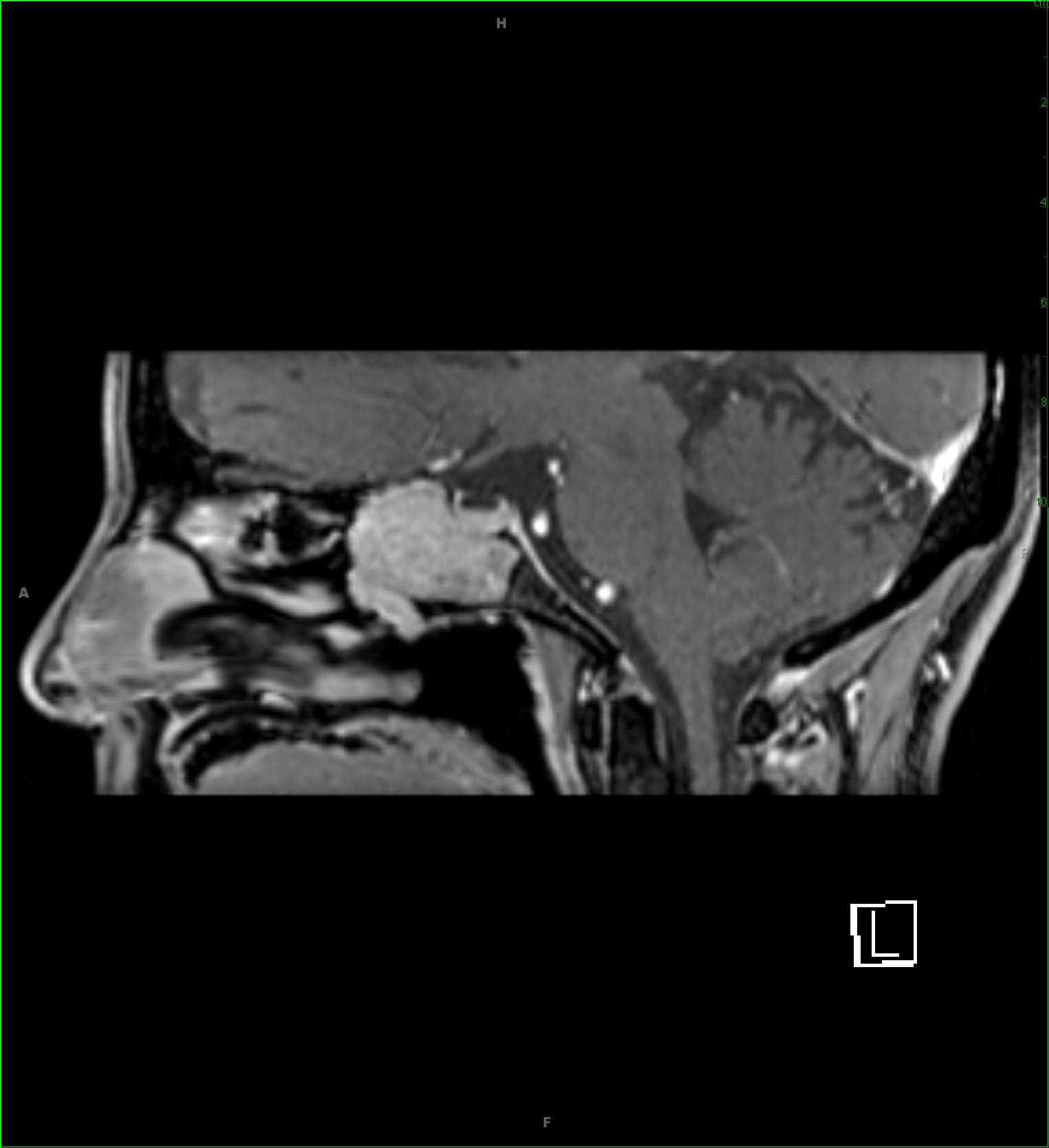
There is a T1-hypointense, T2/STIR-hyperintense, enhancing mass which restricts diffuse at the skull base. The mass involves the clivus, extending from the occipital condyles towards the basisphenoid. The lesion partially effaces the proximal right internal vein at the level of the right jugular foramen. The mass extends into the prepontine and premedullary spaces with partial encasement and leftward displacement of the basilar artery. The imaging findings are compatible with a clival chordoma. Chordomas originate from embryonic remnants of the primitive notochord which extends from the level of Rathke’s pouch to the coccyx. Chordomas are extradural in location, and lytic appearing on CT. Skull base lesions occur at a younger age 30-40 years compared to sacral lesions 50-60 years. Sacrococcygeal lesions are most common, followed by the skull base and vertebral body.
THIS IS CASE
245
OF
396


