- 2
- ,
- 2
- 4
- 6
To Quiz Yourself: Select OFF by clicking the button to hide the diagnosis & additional resources under the case.
Quick Browser: Select ON by clicking the button to hide the additional resources for faster case review.
CASE NUMBER
148


Diagnosis
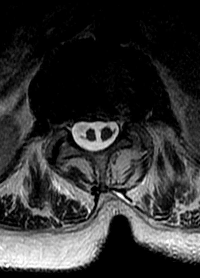
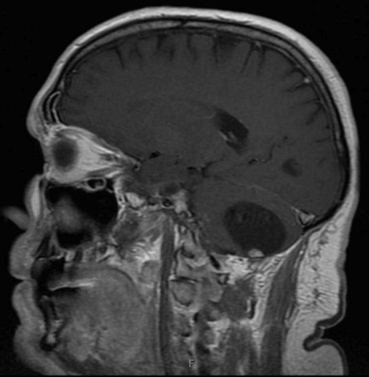
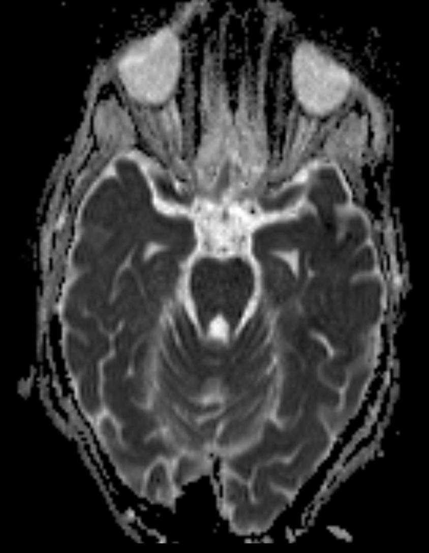
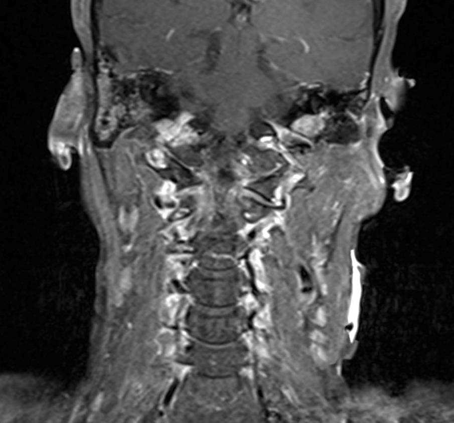
Glomus Jugulare
Note
56-year-old female who presented for dizziness. There are soft tissue masses centered within the jugular foramen on both the left and right. The lesions demonstrate isointense signal on the pre contrast CISS image with heterogenous mildly hyperintense signal on the axial STIR image. Several hypointense foci within the lesions giving them a characteristic pepper appearance. On the noncontrast coronal T1 weighted image, the lesions also demonstrate several hyperintense foci giving them a characteristic salt appearance. The lesions demonstrate mild, heterogeneous enhancement on the axial postcontrast CISS image. Avid enhancement is documented on the postcontrast axial and coronal T1 weighted images. This is a case of bilateral gloms jugulare paraganglioma. The lesion on the right enlarges and occludes the jugular foramen with extension towards the carotid canal. The lesion on the left partially effaces the jugular foramen. The high velocity flow voids within the lesion account for the pepper appearance, while the salt appearance is accounted for by foci of hemorrhage or slow flow within the lesion. Arterial supply is usually via the ascending pharyngeal artery, a branch of the external carotid artery.
Related videos to the case


THIS IS CASE
148
OF
373