- 3
- 5
- ,
- 8
- 7
- 0
To Quiz Yourself: Select OFF by clicking the button to hide the diagnosis & additional resources under the case.
Quick Browser: Select ON by clicking the button to hide the additional resources for faster case review.
CASE NUMBER
4,673


Diagnosis
Giant Cell Tumor Humerus
Note
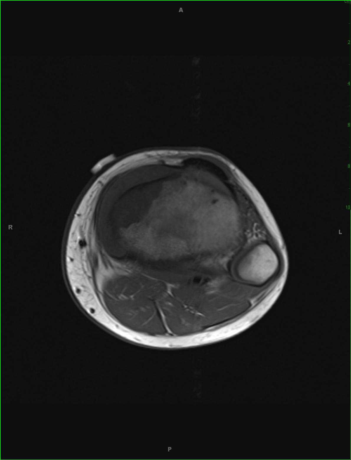
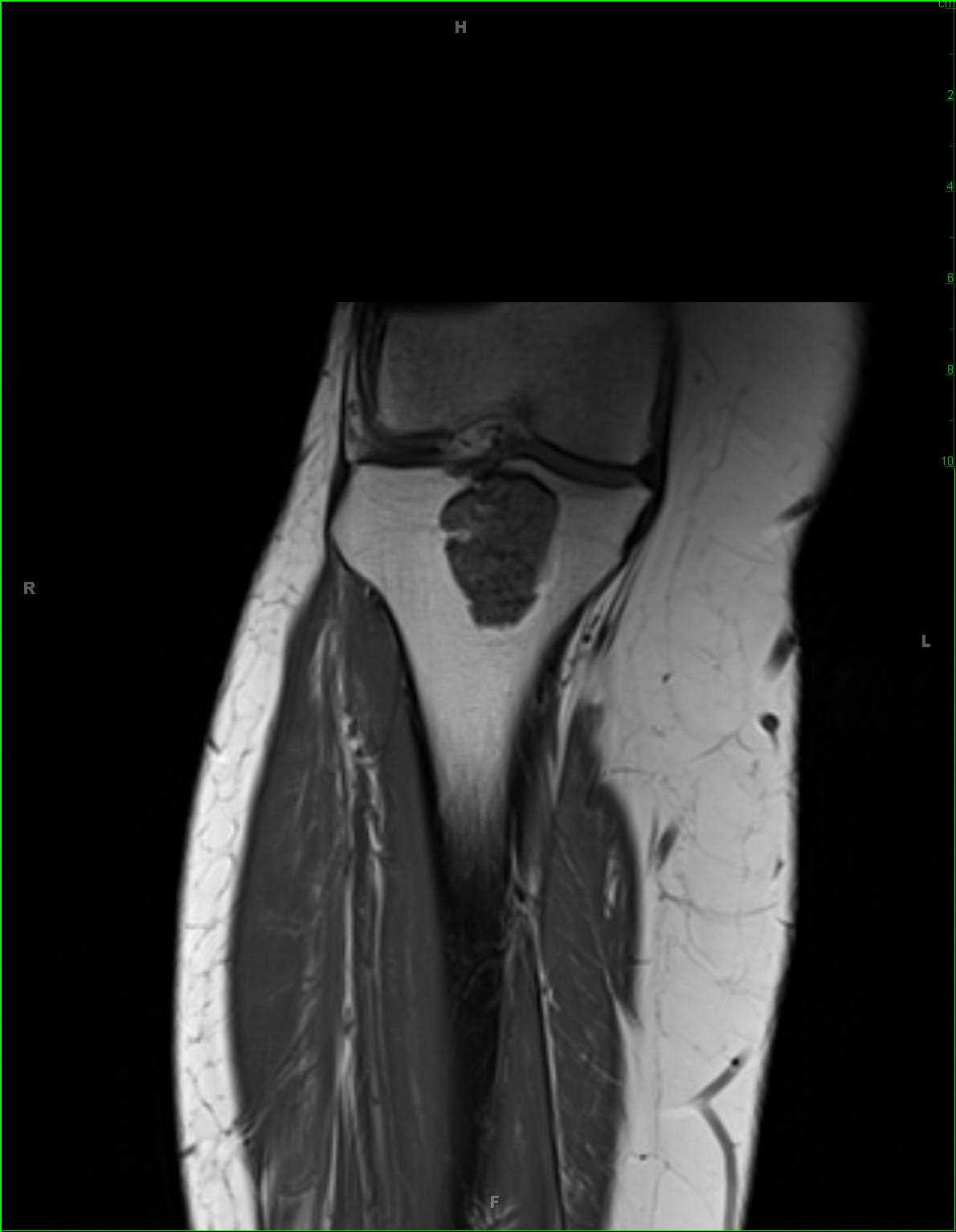
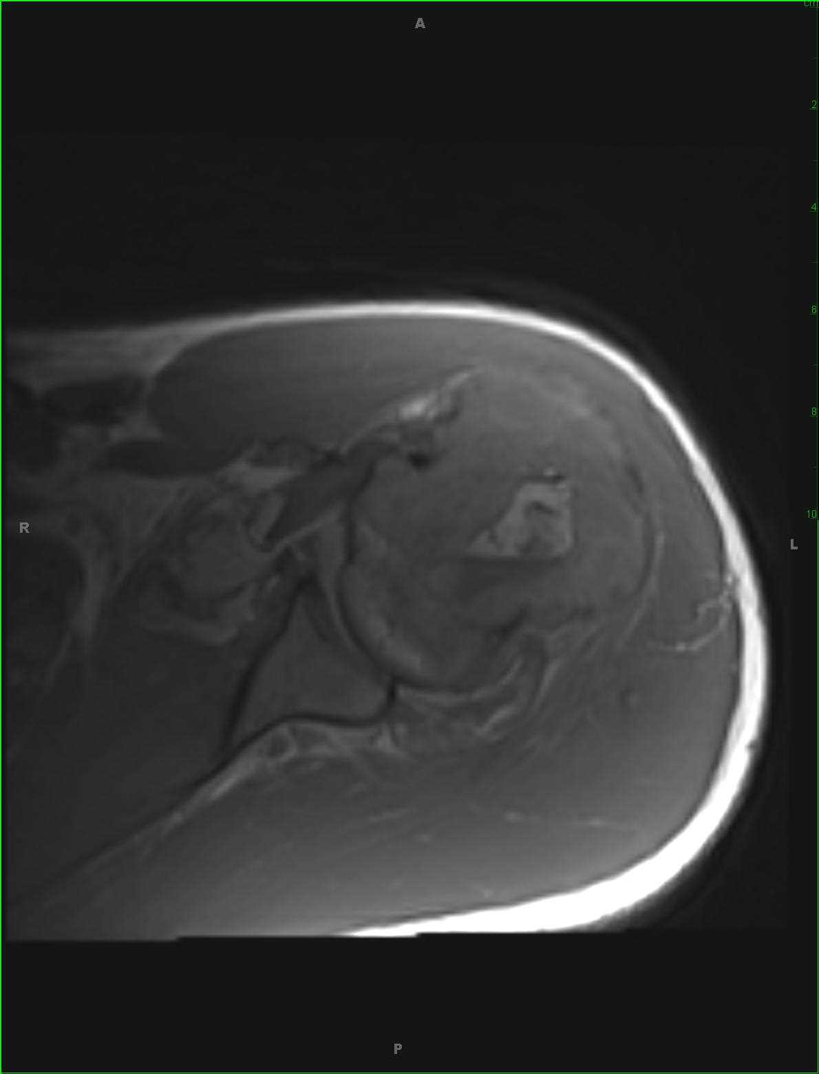
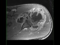
53-year-old female who presents for chronic left shoulder pain. There is an infiltrative proton density isointense, STIR hyperintense, diffusion restricting and heterogeneously enhancing mass involving the proximal left humerus. There is a complex left shoulder effusion with heterogeneous enhancement. The tumor extends to the proximal left humeral epiphysis. Differential diagnosis includes giant cell tumor, chondroblastoma, chondromyxoid fibroma, metastatic disease and plasmacytoma. On biopsy, this was found to be a giant cell tumor. Giant cell tumors are relatively common benign tumors which are found arising from the metaphyseal regions with extension to the physis of long bones. The peak between 20-30 years of age with a slight female predilection. Presentation is usually insidious and related bone pain, soft tissue mass, compression of surrounding structures and possibly pathologic fracture. Lesions more commonly occur in the distal femur, proximal tibia, distal radius, sacrum and vertebral bodies. Treatment is with curettage and packing.
Related videos to the case

THIS IS CASE
4,673
OF
5,587