- 2
- ,
- 3
- 2
- 7
To Quiz Yourself: Select OFF by clicking the button to hide the diagnosis & additional resources under the case.
Quick Browser: Select ON by clicking the button to hide the additional resources for faster case review.
CASE NUMBER
78


Diagnosis
Exophytic hepatic hemangioma
Note
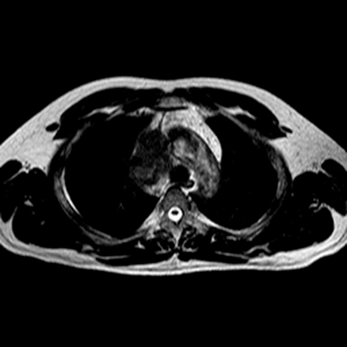
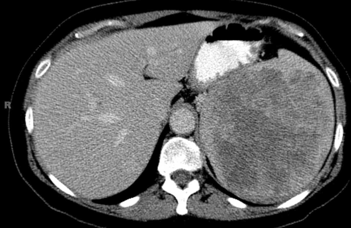
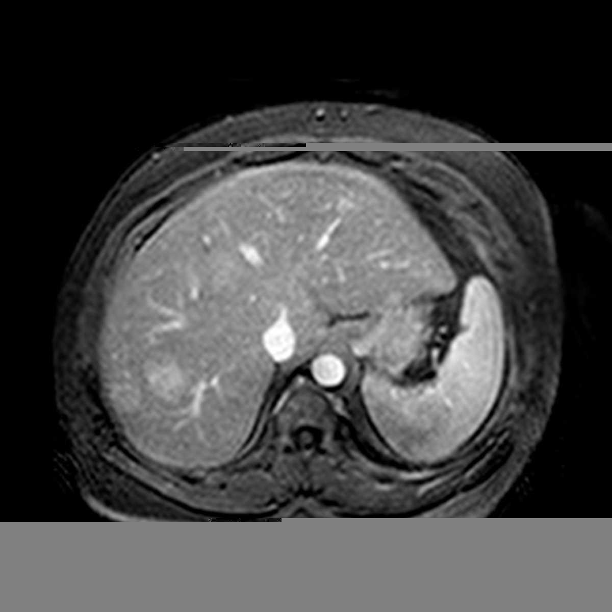
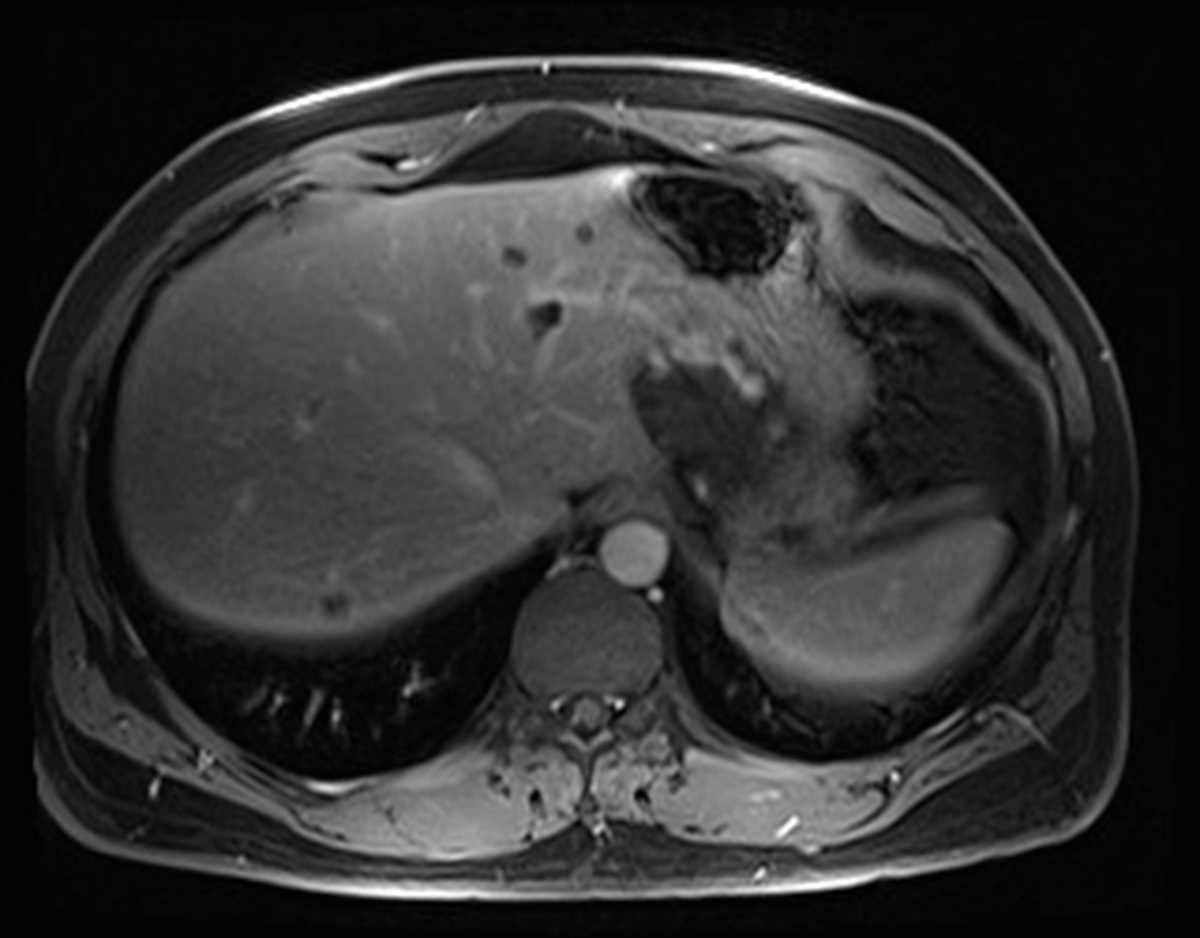
MRI and CT images demonstrate a mass between the liver and stomach which was originally suspected to represent a GIST tumor arising from the stomach. However, the mass is T2 bright, has gradual peripheral globular enhancement on the post-gadolinium images, and appears connected to the liver, in keeping with an unusual exophytic hepatic hemangioma.
Related videos to the case