- 2
- ,
- 2
- 4
- 6
To Quiz Yourself: Select OFF by clicking the button to hide the diagnosis & additional resources under the case.
Quick Browser: Select ON by clicking the button to hide the additional resources for faster case review.
CASE NUMBER
263


Diagnosis
Fibrous Dysplasia, Left Frontal Bone
Note
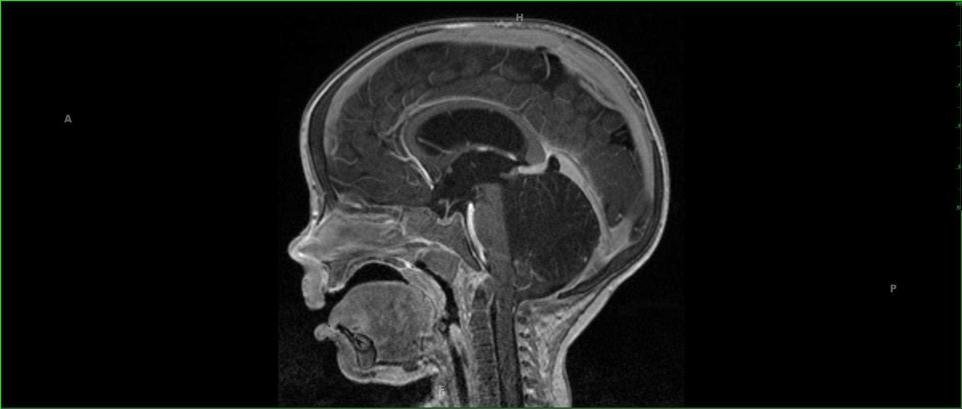
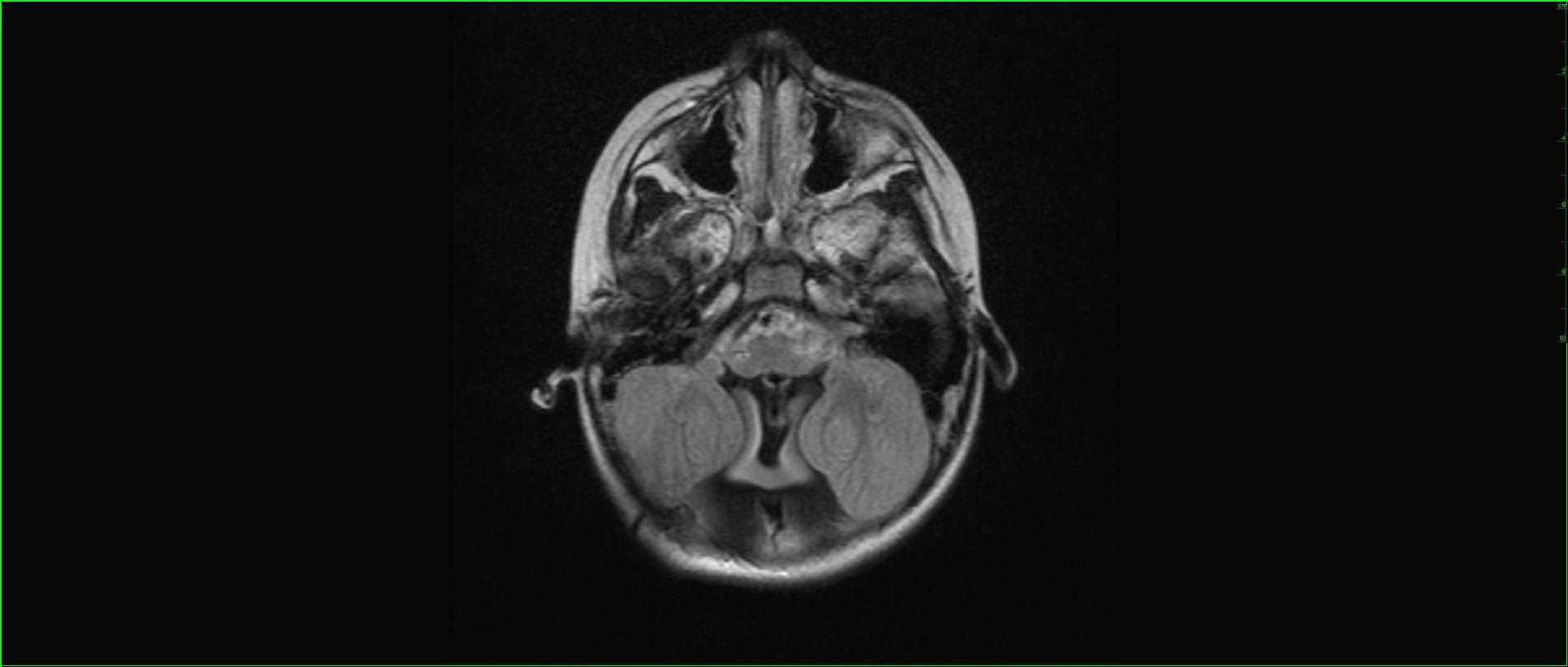
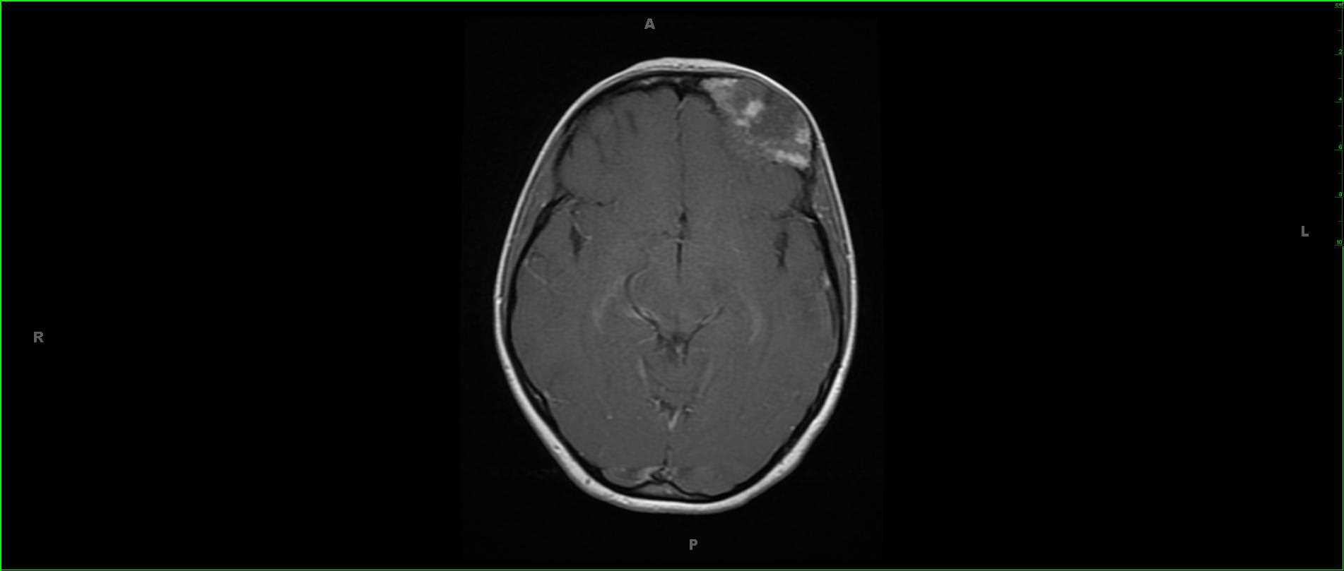
35-year-old female presenting for evaluation of facial asymmetry. Centered within the left frontal bone, there is an expansile, T1 hypointense, T2 hypointense, heterogeneously enhancing lesion with both FLAIR hypointense and slightly hyperintense components which demonstrate mild diffusion restriction. The lesion expands the diploic space of the left frontal bone with extension along the roof of the left orbit. There is no invasion of the underlying dural surface or extraocular musculature. Imaging findings are classic for craniofacial fibrous dysplasia. Typical presentations include cranial asymmetry, facial deformity, nasal stuffiness, proptosis and/or visual impairment. The anterior craniofacial structures are more frequently involved. Heterogeneous signal predominates in the T1 and T2 weighted images. Treatment is typically for cases where the airway or orbital structures are compromised.
Related videos to the case


THIS IS CASE
263
OF
373