- 2
- ,
- 3
- 8
- 1
To Quiz Yourself: Select OFF by clicking the button to hide the diagnosis & additional resources under the case.
Quick Browser: Select ON by clicking the button to hide the additional resources for faster case review.
CASE NUMBER
191


Diagnosis
Multiple Sclerosis
Note
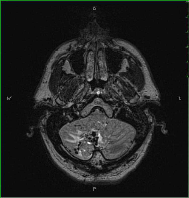
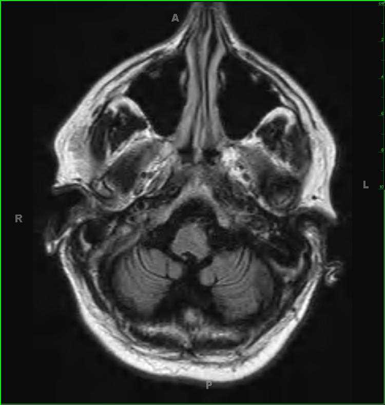
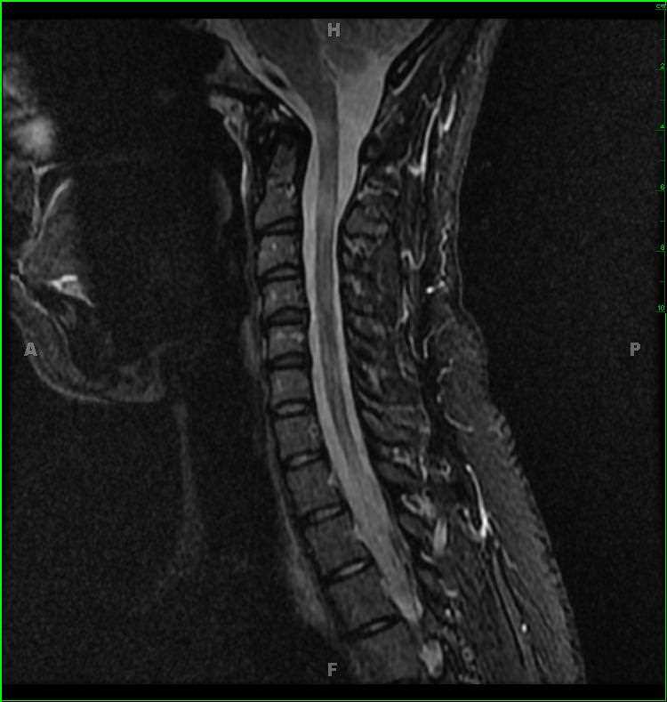
36-year-old female with history of waxing and waning upper and lower extremity parasthesias and weakness of many years duration. There are numerous T2/FLAIR hyperintense lesions within both the supra- and infratentorial compartments, as well as the cervical and upper thoracic spinal region. Ovoid lesions involve the undersurface of the body and splenium of the corpus callosum, as well as the juxtacortical, periventricular, and deep white matter of the brain, brainstem, and cerebellar hemispheres. The supratentorial lesions are oriented perpendicular to the longitudinal axes of the lateral ventricles, in the Dawson fingers configuration. On the axial T2-weighted image, the lesion involving the left parietal centrum semiovale appears as a hole in the white matter. There are numerous lesions in the cervical and upper thoracic spinal regions. The findings are most consistent with a diagnosis of multiple sclerosis. The differential diagnosis includes acute disseminated encephalomyelitis, neuromyelitis optica, and autoimmune mediated vasculitis. Multiple sclerosis is the most common disabling CNS disease of young adults, affecting up to 2.5 million people worldwide. Peak age of onset is 30, with female to male ratio of 2:1.
Related videos to the case


THIS IS CASE
191
OF
396