page 1 | 2 | 3 | 4 | 5 | 6 | 7 | 8 | 9 | 10
Anomalous Anatomy
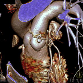
LCA Fistula
Fistula of left main coronary artery and ramus intermedius artery.
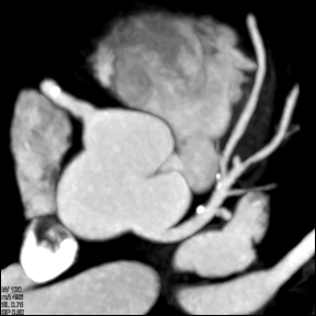
Axial volume rendered shows left main coronary artery coming off the left cusp and dividing into the left anterior descending coronary artery, circumflex coronary artery, and a ramus intermedius branch.
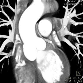
Volume rendered CT shows left main coronary artery coming off the left cusp and a small anomalous vessel between the aorta and the main pulmonary artery.
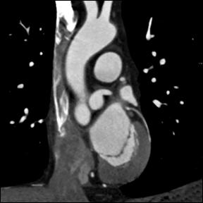
Volume rendered CT shows a normal caliber left main coronary artery and circumflex coronary artery.
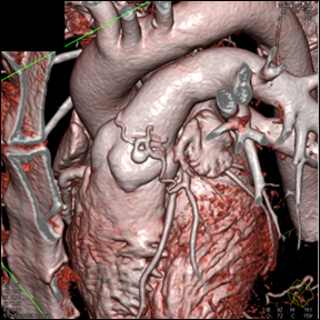
Left main coronary gives rise to a tortuous serpginous vessel that communicates with the main pulmonary artery. A focal aneurysm of the anomalous branch can also seen on the surface of the pulmonary artery as the vessel tracts around the left lateral side of the pulmonary artery.
Left main coronary gives rise to a tortuous serpginous vessel that communicates with the main pulmonary artery. A focal aneurysm of the anomalous branch can also seen on the surface of the pulmonary artery as the vessel tracts around the left lateral side of the pulmonary artery.