- 2
- ,
- 3
- 8
- 1
To Quiz Yourself: Select OFF by clicking the button to hide the diagnosis & additional resources under the case.
Quick Browser: Select ON by clicking the button to hide the additional resources for faster case review.
CASE NUMBER
393


Diagnosis
Meningoencephalocele
Note
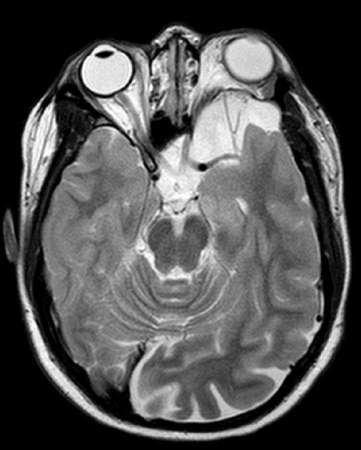
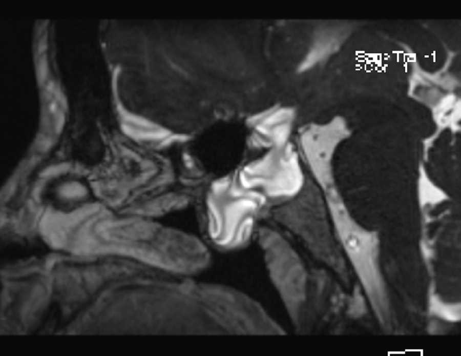
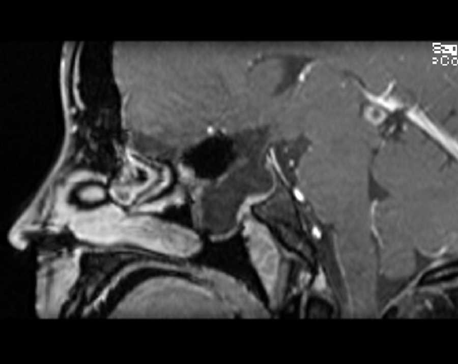
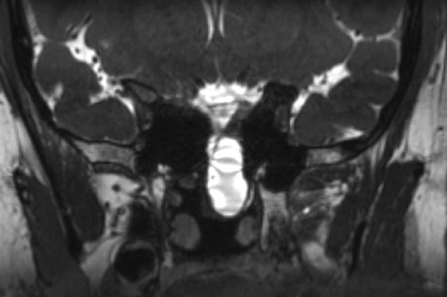
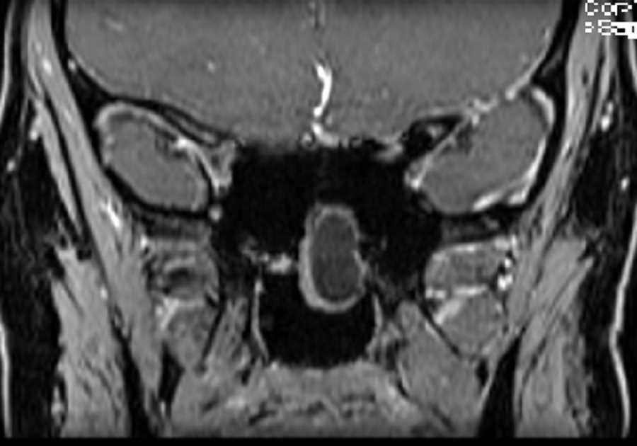
These images demonstrate a large defect in the anterior sella turcica with protrusion of meninges and CSF into the sphenoid sinus and nasopharynx. The optic chiasm and pituitary tissue are also inferiorly displaced and there is marked thinning of the pituitary infundibulum. There is a more subtle defect in the cribiform plate which contains a small portion of the gyrus rectus. No CSF leak was identified Findings are compatible with a meningocele. These lesions can be occult or come to clinical attention by symptoms related to CSF leak. This patient had headaches and endocrinologic disturbances which prompted pituitary imaging.
Related videos to the case

THIS IS CASE
393
OF
396