google ads

Small Bowel Gastrointestinal Stromal Tumors: Imaging and Clinical Correlates

Small Bowel Gastrointestinal Stromal Tumors: Imaging and Clinical Correlates

Franco Verde MD


Disclosures

  • FV: None
  • HR: Royalties, Myriad Genetics, Inc
  • EF: Research support, Siemens AG Advisory Board, Siemens AG Research support, GE Company Advisory Board, HipGraphics, Inc co-founder


Outline of Presentation

  • Introduction
    • Epidemiology
    • Presentation
  • Pathology
    • Terminology
    • Assessment
    • Staging
  • Imaging Appearance
    • CT protocol
    • Location
    • Size
    • Enhancement
    • Additional Features
    • Metastatic Disease
    • Additional cases per site
  • Treatment
  • Prognosis


Introduction: Epidemiology

  • Primary small bowel (SB) tumors are rare
    • 9410 new cases, 1260 deaths (0.2% of all cancer deaths)
  • Gastrointestinal stromal tumors (GISTs) comprise 8-15% of all SB tumors
  • Small bowel GISTs (SB-GISTs) are frequently
    • Older (mean age 60.6 years)
    • Caucasian (79%)
    • Male (55%)


Introduction: Presentation

  • Patients may present with
    • Abdominal pain
    • Bleeding
    • Obstruction
    • Palpable abdominal mass
  • SB-GISTs may also be incidental
    • Frequently when lesion is less than 2 cm
    • When resecting bowel for a different reason (i.e. Whipple procedure for pancreatic adenocarcinoma with tiny incidental duodenal GIST)


Histopathology: History

  • SB-GISTs were previously described as GI leiomyomas, leiomyoblastomas, leiomyosarcomas or neurofibromas or schwannomas
  • Mazar and Clark first used the term gastric stromal tumor in 1983 to describe non-epithelial gastric masses that lacked features of schwann cells or smooth muscle


Pathology: Terminology

  • Originate from common precursor of interstitial cells of Cajal
    • Named after 19th century Spanish neuroanatomist
    • Intermediates between GI autonomic nervous system and smooth muscle cells
    • Found throughout the GI tract around the myenteric plexus in the muscularis propria
    • Kit and Kit-ligand (stem cell factor) positive and dependent cells
      • Kit or Kit-ligand deficient mice lack Cajal cells and have intestinal dysmotility


Pathology: Gross

  • 5 main gross pathologic patterns of SB-GISTs
    • sessile intraluminal small polyps
    • tumors with a small intraluminal and a larger extra-luminal component
    • pedunculated extra-luminal mass
    • extra-luminal mass only
    • large extra-luminal masses with cavitation, hemorrhage, necrosis and luminal communication.
  • Usually pink-tan and occasionally gray-white or yellowish


Pathology: Immunohistology

  • GISTs are predominately related to gain-of-function mutations of KIT gene
    • Which encodes for tyrosine kinase transmembrane receptor
    • 85% of all GISTs have some variable mutation of the KIT gene: typically exons 9, 11, 13, or 17
    • Each exon codes for a different component of the tyrosine receptor
    • Receptor responsible for initiating a signaling cascade involved in cellular survival, growth, and differentiation
  • Up to 10% of GISTs without detectable KIT gene mutation will have a PDGRFA gene, another tyrosine kinase cell surface receptor.
  • Rarely, no KIT or PDGRFA gene mutation is found. Mutations have been found in other downstream genes (e.g. BRAF, NF1, or RAS)


Pathology: Assessment

Modern pathologic assessment of GISTs include the following parameters
  • Location
  • Size
  • Subtype
  • # of mitoses per 50 high powered fields (HPFs)
  • Presence of necrosis
  • Presence of lymphatic and venous invasion
  • Surgical margin assessment
  • Node assessment
  • Presence of metastatic disease
  • Immunostains
  • Preoperative treatment


Pathology: Assessment

  • Location
    • Gastric, small bowel, etc.
    • Small bowel GISTs have been shown to have a higher rate of progressive disease compared to gastric origin
    • Duodenal GISTs are more common than jejunal or ileal GISTs
  • Size
    • Main determinant in prognosis and TNM classification
    • Categorized into
      • T1: < 2 cm
      • T2: > 2 cm and < 5 cm
      • T3: > 5 cm and < 10 cm
      • T4: > 10 cm


Pathology: Assessment

  • Subtype
    • Spindle cell morphology is most common
    • Small percentage epithelioid
    • Rarely mixed pattern
  • Mitotic Rate
    • Next most important determinant of risk of progressive disease
    • G1 or low-grade: less than 5 mitoses per 50 HPFs
    • G2 or high-grade: more than 5 mitoses per 50 HPFs
  • Necrosis
    • Either coagulative or liquefactive
    • Presence of necrosis is not a determinant of progression
  • Lymphatic or venous invasion
    • Described but not used in staging


Pathology: Assessment

  • Nodal disease
    • Rarely does GIST metastasize to regional nodes
  • Metastatic disease
    • Found as peritoneal implants or hepatic disease
  • Immunostains
    • Positive and routinely tested for evaluating a small bowel tumor:
      • C-kit
      • DOG-1 (cell-surface novel marker, not usually found in other soft tissue tumors)
      • PDGFRA (tested when C-kit is negative)
    • Negative and routinely tested for:
      • Desmin and SMA – smooth muscle markers found in leiomyomas
    • S100, HMB45, cytokeratin – found in tumors of epidermal origin (e.g. schwannoma or melanoma)
  • Preoperative treatment: if a tyrosine receptor blocker was used (i.e. imatinib mesylate)


Pathology: AJCC Staging

Pathology:AJCC Staging


Imaging: Protocol Optimization

Recommended 64 MDCT protocol
  • Arterial (bolus triggered) and venous phases (60 second delay) at 4-5 mL/s
  • 120 kVp
  • 250 mAs
  • 0.8 pitch
  • 64 x 0.6 mm collimation
  • 0.75 mm axial and 3 mm axial, sagittal, and coronal reconstructions
  • 1000 mL water PO


Imaging: Location

  • Typically appear as extra-luminal masses with a small intra-luminal component (tip-of-iceberg)
  • Duodenum more common than jejunum or ileum
  • Example of a low grade, 1.9 cm, spindle type GIST arising from the duodenum with tiny intraluminal component (arrow)
Imaging:Location


Imaging: Location

  • Duodenal GISTs can mimic neuroendocrine tumors given seemingly completely extraluminal appearance
  • Example of a 3.6 cm a low grade, spindle type, GIST arising from the duodenum.
    • Mass was resected with pre-operative diagnosis of neuroendocrine tumor
Imaging:Location


Imaging: Location

  • Some very large SB GISTs may have no apparent source and seemingly arise from the mesentery and mimic other sarcomas
  • Example of 33 cm high grade jejunal GIST with 50% necrosis with extensive local mass effect
Imaging:Location


Imaging: Size

  • SB GISTs demonstrate a spectrum of sizes typically 5 to 10 cm with approximate similar percentages of less than 5 cm or greater than 10 cm
  • Example of a 5 cm low grade jejunal GIST
Imaging:Location


Imaging: Enhancement

  • Noncontrast demonstrates homogenous soft tissue density (lower arrow)
  • Focal calcifications can be seen (upper arrow)
  • Example of a 6.5 cm high grade, necrotic, GIST arising from distal jejunum.
Imaging:Location


Imaging: Enhancement

  • Enhancement pattern is variable depending on size, phase of contrast, and presence of necrosis or luminal connection
  • Example of a 2.5 cm duodenal GIST with heterogeneous enhancement on arterial (A) phase becoming homogenous on venous (B) phase. Note degree of enhancement is similar to renal cortex
Imaging:Enhancement


Imaging: Enhancement

  • If the GIST is large, necrosis will likely be present and appear and central hypoattenuation and a rim of enhancing viable tissue.
    • Example of a high grade duodenal GIST with extensive necrosis.
  • Central heterogeneous enhancement in smaller GISTs (lower image) does not necessarily indicate necrosis.
    • Example of a low grade duodenal GIST without necrosis.
Imaging:Enhancement


Imaging: Additional Features

  • Intratumoral air and fluid levels or debris can be seen when there is luminal communication
  • Example of low grade GIST arising from the duodenum with extensive necrosis.
    • Note luminal communication demonstrated by intratumoral air (arrow)
Imaging:Additional Features


Imaging: Metastatic Disease

  • Metastatic disease is usually seen as mesenteric or liver lesions
  • Mesenteric nodules are usually small, round and may enhance.
  • Liver metastases are hypovascular, round, and well demarcated.
Imaging:Metastatic Disease


Imaging Additional Cases - Duodenum

ImagingAdditional Cases - Duodenum


Imaging Additional Cases - Jejunum

ImagingAdditional Cases - Jejunum


Imaging: Additional Cases - Ileum

Imaging:Additional Cases - Ileum


Treatment

  • Surgical resection is mainstay therapy for isolated small tumors without evidence of metastatic disease.
  • Laparoscopic resection may be attempted for small tumors with open procedures reserved for larger tumors.
  • Imatinib mesylate, a tyrosine kinase inhibitor, is first line therapy for patients who are poor surgical candidates, or show evidence of recurrence or metastatic disease.
    • Can also be used as neoadjuvant therapy to preoperatively shrink large tumors


Imaging Surveillance

  • Surveillance after surgery includes CT scanning with contrast every 3 to 6 months.
  • Low grade small tumor may have scans every 6 to 12 months.
  • Patients receiving imatinib mesylate therapy may have scans every 3 months.
  • Dual phase imaging is recommended as arterial phase may detect new enhancing components in stable disease that may wash out during venous phase and be overlooked.


Prognosis: Size and Mitotic Rate Dependent

PrognosisSize and Mitotic Rate Dependent


References

  • Anonymous SEER Cancer Statistics Factsheets: Small Intestine Cancer. National Cancer Institute. Bethesda, MD.
    . http://seer.cancer.gov/statfacts/html/smint.html. 08/11. 2015.
  • Bilimoria KY, Bentrem DJ, Wayne JD, Ko CY, Bennett CL, Talamonti MS. Small bowel cancer in the United States: changes in epidemiology, treatment, and survival over the last 20 years. Ann Surg 2009;249(1):63-71.
  • Miettinen M, Lasota J. Gastrointestinal stromal tumors: pathology and prognosis at different sites. Semin Diagn Pathol 2006;23(2):70-83.
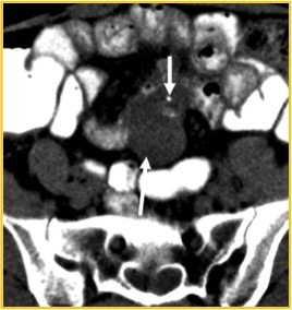
  • Kukar M, Kapil A, Papenfuss W, Groman A, Grobmyer SR, Hochwald SN. Gastrointestinal stromal tumors (GISTs) at uncommon locations: a large population based analysis. J Surg Oncol 2015;111(6):696-701.
  • Miettinen M, Lasota J. Gastrointestinal stromal tumors--definition, clinical, histological, immunohistochemical, and molecular genetic features and differential diagnosis. Virchows Arch 2001;438(1):1-12.
  • Crosby JA, Catton CN, Davis A, et al. Malignant gastrointestinal stromal tumors of the small intestine: a review of 50 cases from a prospective database. Ann Surg Oncol 2001;8(1):50-59.
  • Pidhorecky I, Cheney RT, Kraybill WG, Gibbs JF. Gastrointestinal stromal tumors: current diagnosis, biologic behavior, and management. Ann Surg Oncol 2000;7(9):705-712.
  • DeMatteo RP, Lewis JJ, Leung D, Mudan SS, Woodruff JM, Brennan MF. Two hundred gastrointestinal stromal tumors: recurrence patterns and prognostic factors for survival. Ann Surg 2000;231(1):51-58.
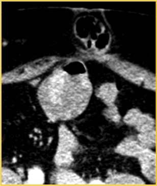
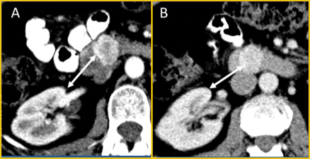
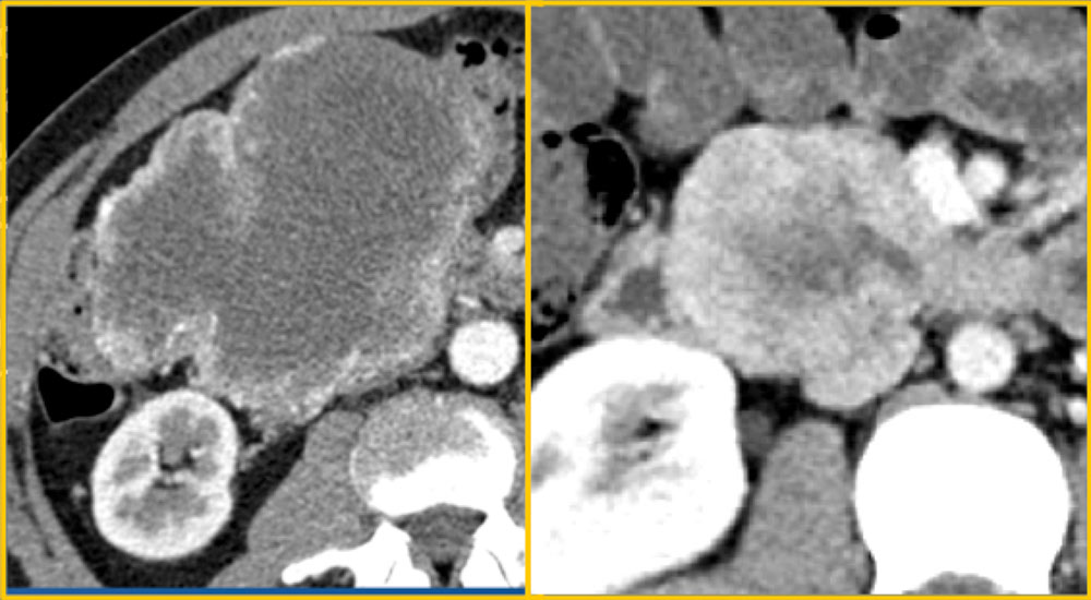
  • Grover S, Ashley SW, Raut CP. Small intestine gastrointestinal stromal tumors. Curr Opin Gastroenterol 2012;28(2):113-123.
  • Kimura H, Yoshida T, Kinoshita S, Takahashi I. Pedunculated giant gastrointestinal stromal tumor of the stomach showing extragastric growth: report of a case. Surg Today 2004;34(2):159-162.
  • Miettinen M, Fetsch JF, Sobin LH, Lasota J. Gastrointestinal stromal tumors in patients with neurofibromatosis 1: a clinicopathologic and molecular genetic study of 45 cases. Am J Surg Pathol 2006;30(1):90-96.
  • Andersson J, Sihto H, Meis-Kindblom JM, Joensuu H, Nupponen N, Kindblom LG. NF1-associated gastrointestinal stromal tumors have unique clinical, phenotypic, and genotypic characteristics. Am J Surg Pathol 2005;29(9):1170-1176.
  • Ozcinar B, Aksakal N, Agcaoglu O, et al. Multiple gastrointestinal stromal tumors and pheochromocytoma in a patient with von Recklinghausen's disease. Int J Surg Case Rep 2013;4(2):216-218.
  • Miettinen M, Makhlouf H, Sobin LH, Lasota J. Gastrointestinal stromal tumors of the jejunum and ileum: a clinicopathologic, immunohistochemical, and molecular genetic study of 906 cases before imatinib with long-term follow-up. Am J Surg Pathol 2006;30(4):477-489.
  • Hirota S, Isozaki K, Moriyama Y, et al. Gain-of-function mutations of c-kit in human gastrointestinal stromal tumors. Science 1998;279(5350):577-580.
  • Joensuu H, Hohenberger P, Corless CL. Gastrointestinal stromal tumour. Lancet 2013;382(9896):973-983.
  • Lamba G, Ambrale S, Lee B, Gupta R, Rafiyath SM, Liu D. Recent advances and novel agents for gastrointestinal stromal tumor (GIST). J Hematol Oncol 2012;5:21-8722-5-21.
  • Edling CE, Hallberg B. c-Kit--a hematopoietic cell essential receptor tyrosine kinase. Int J Biochem Cell Biol 2007;39(11):1995-1998.
  • Miettinen M, Wang ZF, Lasota J. DOG1 antibody in the differential diagnosis of gastrointestinal stromal tumors: a study of 1840 cases. Am J Surg Pathol 2009;33(9):1401-1408.
  • Miettinen M, Virolainen M, Maarit-Sarlomo-Rikala. Gastrointestinal stromal tumors--value of CD34 antigen in their identification and separation from true leiomyomas and schwannomas. Am J Surg Pathol 1995;19(2):207-216.
  • Wu TJ, Lee LY, Yeh CN, et al. Surgical treatment and prognostic analysis for gastrointestinal stromal tumors (GISTs) of the small intestine: before the era of imatinib mesylate. BMC Gastroenterol 2006;6:29.
  • Nishida T, Blay JY, Hirota S, Kitagawa Y, Kang YK. The standard diagnosis, treatment, and follow-up of gastrointestinal stromal tumors based on guidelines. Gastric Cancer 2015
  • Fletcher CD, Berman JJ, Corless C, et al. Diagnosis of gastrointestinal stromal tumors: A consensus approach. Hum Pathol 2002;33(5):459-465.
  • Jang SH, Kwon JE, Kim JH, et al. Prediction of Tumor Recurrence in Patients with Non-Gastric Gastrointestinal Stromal Tumors Following Resection according to the Modified National Institutes of Health Criteria. Intest Res 2014;12(3):229-235.

Acknowldgements

  • Franco Verde MD
  • Ralph Hruban MD
  • Elliot K. Fishman MD

Privacy Policy

Copyright © 2024 The Johns Hopkins University, The Johns Hopkins Hospital, and The Johns Hopkins Health System Corporation. All rights reserved.