
Multidisciplinary approach for program development with artificial intelligence in pancreatic cancer: How we fit in
Multidisciplinary approach for program development with artificial intelligence in pancreatic cancer: How we fit in Satomi Kawamoto, M.D. The Russell H. Morgan Department of Radiology and Radiological Science, Department of Computer Science, Department of Pathology, and the Department of Cancer Research Johns Hopkins University |
Disclosure Research supported by the Lustgarten Foundation
|
Learning Objectives
|
Introduction (1)
|
Introduction (2)
|
Deep Learning Applications in Cancer Imaging
|
Goals of Deep Learning Models To create mathematical models that can be:
|
Multidisciplinary Approach in Development of AI Algorithm for Detection of Early Pancreatic Cancer Radiologists play a critical role in development of AI in medical imaging as a member of multidisciplinary team 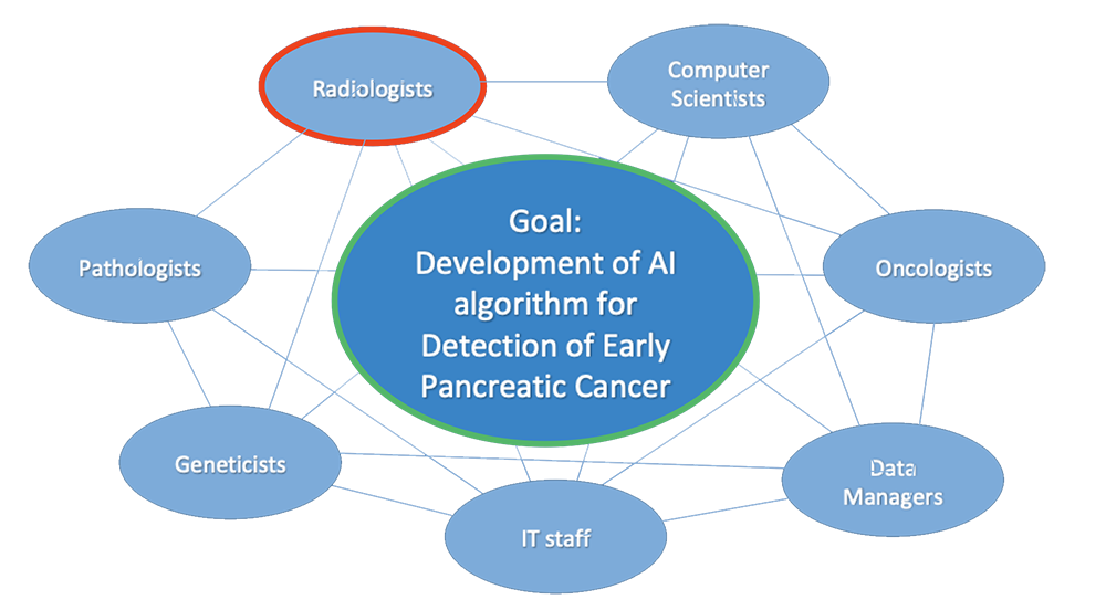 |
Roles of Radiologists in Development of DL algorithms Development of deep learning algorithms for early detection of pancreatic cancer 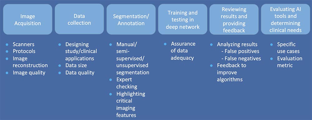 |
Image Acquisition
|
Data Collection
|
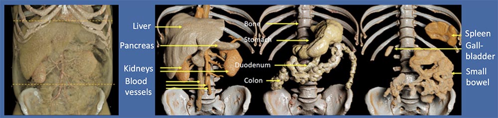
Data Collection/Segmentation Process and Workflow We developed an unique and reliable data collection and annotation process for normal abdominal structures using volumetric CT that can be used to train the deep learning network for automated recognition of normal abdominal organs. 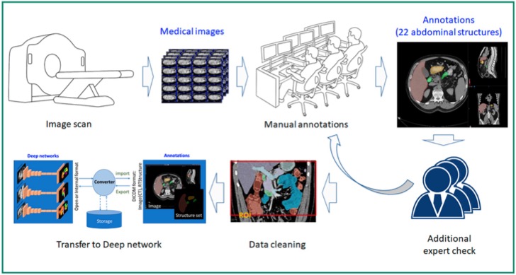 Park S, et al. Annotated normal CT data of the abdomen for deep learning. Diagnostic and Interventional Imaging 2019 [Epub ahead of print] |
Image Segmentation
|
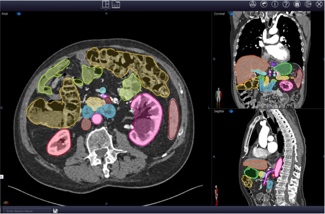
Image Segmentation (Normal CT) Image segmentation: Partitioning an image into multiple regions that share similar attributes, enabling localization and quantification. Manually segmented normal abdominal structures on Coronal CT image 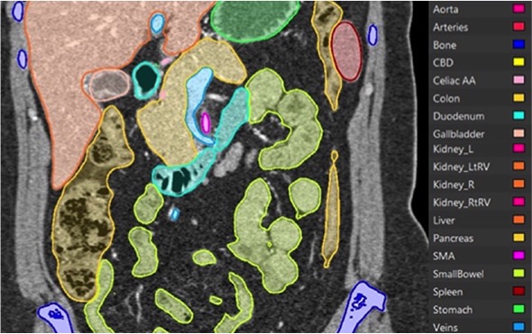 Park S, et al. Diagnostic and Interventional Imaging 2019 [Epub ahead of print] |
2D vs. 3D Image Segmentation
 Park S, et al. Diagnostic and Interventional Imaging 2019 [Epub ahead of print] |
Deep Network Predictions
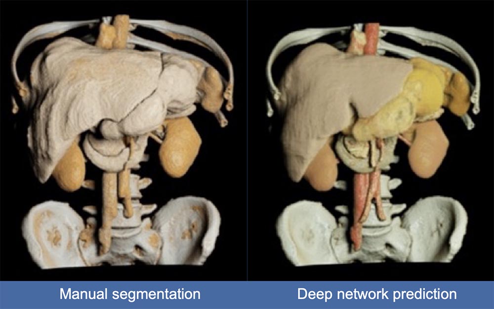 Wang Y, et al. http://arxiv.org/abs/1804.08414 Park S, et al. Diagnostic and Interventional Imaging 2019 [Epub ahead of print] |
Image Segmentation (Pancreatic Cancer)
 |
Supervised vs. Unsupervised Learning (1)
|
Supervised vs. Unsupervised Learning (2)
|
Training and Testing in Deep Network
|
Example of False Negative Case of Deep Network Prediction Pancreatic neuroendocrine tumor in the body of the pancreas was not predicted by deep network. Radiologists review the casesand provide feedback to the multidisciplinary team to improve algorithms. 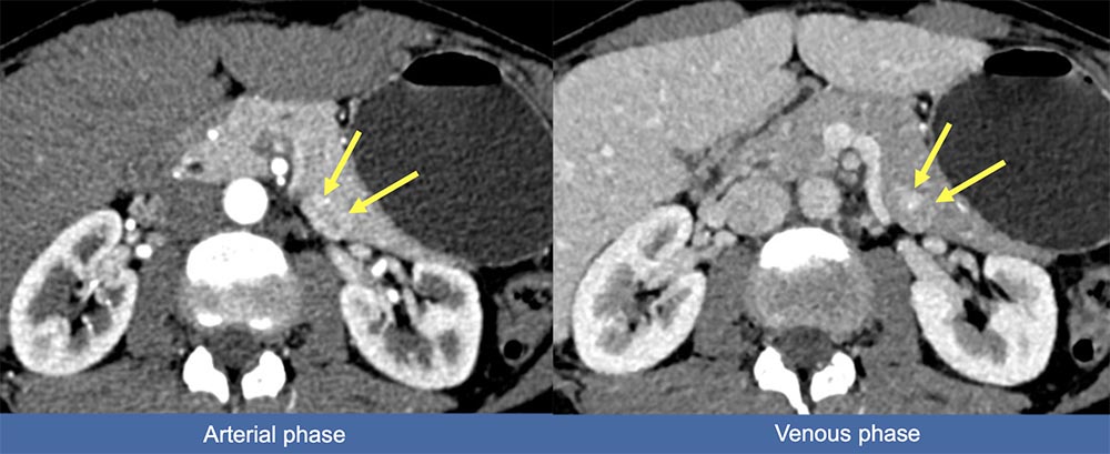 |
Example of False Negative Case of Deep Network Prediction Pancreatic neuroendocrine tumor in the head of the pancreas was not predicted by deep network. Radiologists review the casesand provide feedback to the multidisciplinary team to improve algorithms. 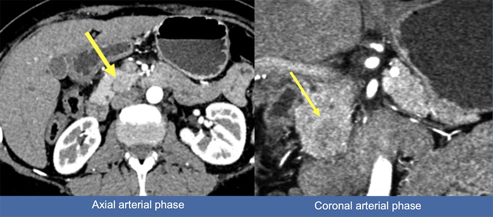 |
Example of False Positive Case of Deep Network Prediction Uneven focal fatty infiltration of the head of the pancreas might be related to false positive prediction False positive PDAC prediction in the head of the pancreas 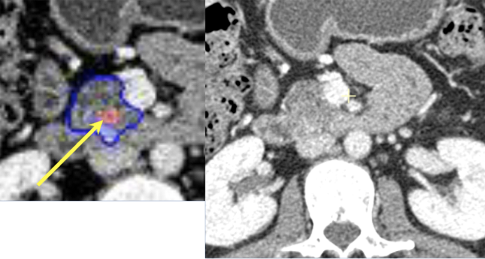 Wang Y, Zhou Y, Shen W, Park S, Fishman EK, Yuille AL. Abdominal multi-organ segmentation with organ-attention networks and statistical fusion. https://arxiv.org/abs/1804.08414 |
Deep Network Prediction for Detection of PNET Found Multiple Tumors: Need Confirmation of Multi-tumor Cases vs. Annotation/Segmentation Error Pancreatic serous cystadenoma (blue arrow) in the head of the pancreas and neuroendocrine tumor (yellow arrow) in the tail of the pancreas. Radiologists confirmed this was a single PNET case with incidental other tumor by reviewing the CT and pathology data. 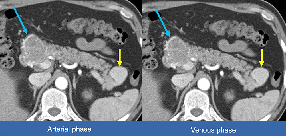 |
Evaluation of AI Tools
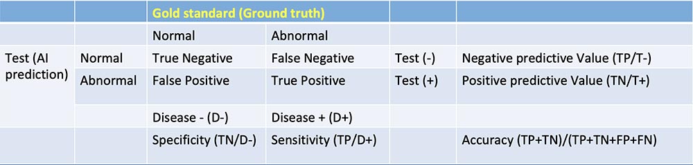 |
Evaluation of AI Tools
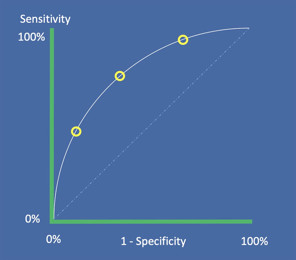 Rubin DL. J Am Coll Radiol 2019;16:1309-1317 |
Determining Clinical Needs
|
Discussion (1)
|
Discussion (2)
|
Conclusion
|
References
Acknowledgements
|