Imaging Pearls ❯ 3D and Workflow ❯ Bone Editing
|
-- OR -- |
|
- Cinematic Rendering: The Reality in Clinical Practice
- Robust rendering technique across multiple body regions ranging from head to feet
- Excellent for demonstration of skin, muscle, soft tissues, bone, vasculature
- Complex trapizoids can allow for multiple tissue types to be displayed in a single visualization - Cinematic Rendering: The Reality in Clinical Practice
- Preset values of specific anatomic values can be used in most cases to speed up the generation of the 3D image dataset
- Preset values may change depending on the specific visualization (anterior vs lateral view) and the scan protocol used (IV contrast and injection rate and acquisition timing such as arterial/venous/delayed phase imaging) - Cinematic Rendering: The Reality in Clinical Practice
- AI may prove to be of value in choosing the best (or a series of potential best) image displays based on the dataset used.
- For example could the system provide a series of select “preset views” to image a suspected pancreatic mass based on learning from previously generated datasets? - Cinematic Rendering: The Reality in Clinical Practice
- Visualization of cinematic rendered images can be done with select images or with the creation of a video file. Video files have the advantage over static images especially for the referring physician who may not have access to the workstation
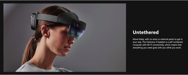
- Newer display technologies like Microsoft HoloLens 2 may enhance the value of cinematic rendering - HoloLens 2
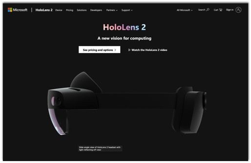
- HoloLens 2

- HoloLens 2

- HoloLens 2

- HoloLens 2
- "The purpose of this manuscript is to describe a promising automated segmentation tool, which uses and extends an Interactive Watershed Transform (IWT) technique to remove selected portions of the anatomy."
Automated Multidetector Row CT Dataset Segmentation with an Interactive Watershed Transform (IWT) Algorithm:Part 1
Understanding the IWT Technique
Heath DG, Hahn HK, Johnson PT, Fishman EK
J Digital Imaging (in press) - "The purpose of this paper is to demonstrate the utility of a fast, automated editing tool, an Interactive Watershed Transform (IWT) technique, for segmenting body multidetector row computed tomography (MDCT) volumes."
Automated Multidetector Row CT Dataset Segmentation with an Interactive Watershed Transform (IWT) Algorithm:Part 2-Body CT Angiographic and Orthopedic Applications
Heath DG, Hahn HK, Johnson PT, Fishman EK
J Digital Imaging (in press) - "Selective removal of bones enhances 3D volume-rendered viewing of surfaces, particularly in joint articulations.The information provided from surface viewing is valuable for both diagnosis and presurgical planning in the setting of arthropathy and fracture."
Automated Multidetector Row CT Dataset Segmentation with an Interactive Watershed Transform (IWT) Algorithm:Part 2-Body CT Angiographic and Orthopedic Applications
Heath DG, Hahn HK, Johnson PT, Fishman EK
J Digital Imaging (in press) - "Whereas editing with IWT is an efficient method of improving the diagnostic capacity of 3D-rendered imaging,errors can still arise when vessels are located in close proximity to bone."
Automated Multidetector Row CT Dataset Segmentation with an Interactive Watershed Transform (IWT) Algorithm:Part 2-Body CT Angiographic and Orthopedic Applications
Heath DG, Hahn HK, Johnson PT, Fishman EK
J Digital Imaging (in press)